
Page 519 - Physics Coursebook 2015 (A level)
P. 519
Chapter 32: Medical imaging
Applying physics
In this book, you have learned many important ideas from physics. You may have noticed that the same big ideas keep reappearing – for example, the idea of a field of force (magnetic, electric, gravitational), or the idea of energy transmitted as waves, or the idea that matter is made of particles with forces acting between them. This is an important characteristic of physics; ideas that are used in one area prove to be useful in another. Hopefully, you will see many of these connections now that you are approaching the end of your course.
Physics is also useful. It is applied in many
areas of life. In this chapter, we look at one of these areas: medical imaging. This topic covers a range
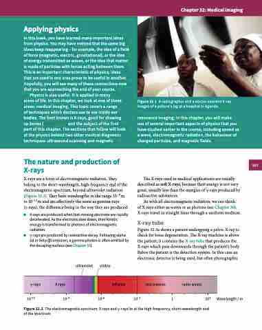
of techniques which doctors use to see inside our bodies. The best known is X-rays, good for showing up bones (Figure 32.1), and the subject of the first part of this chapter. The sections that follow will look at the physics behind two other medical diagnostic techniques: ultrasound scanning and magnetic
Figure 32.1 A radiographer and a doctor examine X-ray images of a patient’s leg at a hospital in Uganda.
resonance imaging. In this chapter, you will make use of several important aspects of physics that you have studied earlier in the course, including sound as a wave, electromagnetic radiation, the behaviour of charged particles, and magnetic fields.
The nature and production of X-rays
X-rays are a form of electromagnetic radiation. They belong to the short-wavelength, high-frequency end of the electromagnetic spectrum, beyond ultraviolet radiation (Figure 32.2). They have wavelengths in the range 10−8 m to 10−13 m and are effectively the same as gamma-rays (γ-rays), the difference being in the way they are produced:
■■ X-rays are produced when fast-moving electrons are rapidly decelerated. As the electrons slow down, their kinetic energy is transformed to photons of electromagnetic radiation.
■■ γ-rays are produced by radioactive decay. Following alpha (α) or beta (β) emission, a gamma photon is often emitted by the decaying nucleus (see Chapter 16).
The X-rays used in medical applications are usually described as soft X-rays, because their energy is not very great, usually less than the energies of γ-rays produced by radioactive substances.
As with all electromagnetic radiation, we can think
of X-rays either as waves or as photons (see Chapter 30). X-rays travel in straight lines through a uniform medium.
X-ray tube
Figure 32.3a shows a patient undergoing a pelvic X-ray to check for bone degeneration. The X-ray machine is above the patient; it contains the X-ray tube that produces the X-rays which pass downwards through the patient’s body. Below the patient is the detection system. In this case an electronic detector is being used, but often photographic
ultraviolet
visible
10–6 10–3
infrared
γ-rays X-rays
10–12 10–9
microwaves
radio waves
Figure 32.2 The electromagnetic spectrum; X-rays and γ-rays lie at the high-frequency, short-wavelength end of the spectrum.
1
103 Wavelength / m
507
