Page 230 - Understanding Psychology
P. 230
   Your Blind
Spot
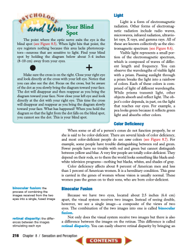
The point where the optic nerve exits the eye is the blind spot (see Figure 8.5). When light hits that point, the eye registers nothing because this area lacks photorecep- tors—neurons that are sensitive to light. Find your blind spot by holding the diagram below about 3–4 inches (8–10 cm•) away from your eyes. +
Make sure the cross is on the right. Close your right eye and look directly at the cross with your left eye. Notice that you can also see the dot. Focus on the cross, but be aware of the dot as you slowly bring the diagram toward your face. The dot will disappear and then reappear as you bring the diagram toward your face. Now close your left eye and look directly at the dot with your right eye. This time the cross will disappear and reappear as you bring the diagram slowly toward your face. What has happened? When you hold the diagram so that the light from the dot falls on the blind spot, you cannot see the dot. This is your blind spot.
   binocular fusion: the process of combining the images received from the two eyes into a single, fused image
retinal disparity: the differ- ences between the images stimulating each eye
Light is a form of electromagnetic radiation. Other forms of electromag- netic radiation include radio waves, microwaves, infrared radiation, ultravio- let rays, X rays, and gamma rays. All of these are known collectively as the elec- tromagnetic spectrum (see Figure 8.6).
Visible light represents a small por- tion of the electromagnetic spectrum, which is composed of waves of differ- ent length and frequency. You can observe the wavelengths of visible light with a prism. Passing sunlight through a prism breaks the light into a rainbow of colors. Each of these colors is com- prised of light of different wavelengths. While prisms transmit light, other objects absorb and reflect light. The ob- ject’s color depends, in part, on the light that reaches our eyes. For example, a pea looks green because it reflects green light and absorbs other colors.
Color Deficiency
When some or all of a person’s cones do not function properly, he or she is said to be color-deficient. There are several kinds of color deficiency, and most color-deficient people do see some colors (see Figure 8.7). For example, some people have trouble distinguishing between red and green. Fewer people have no trouble with red and green but cannot distinguish between yellow and blue. A very few people are totally color-deficient. They depend on their rods, so to them the world looks something like black-and- white television programs—nothing but blacks, whites, and shades of gray.
Color deficiency affects about 8 percent of American men and less than 1 percent of American women. It is a hereditary condition. This gene is carried in the genes of women whose vision is usually normal. These women pass the genes on to their sons, who are born color-deficient.
Binocular Fusion
Because we have two eyes, located about 2.5 inches (6.4 cm) apart, the visual system receives two images. Instead of seeing double, however, we see a single image—a composite of the views of two eyes. The combination of the two images into one is called binocular fusion.
Not only does the visual system receive two images but there is also a difference between the images on the retinas. This difference is called retinal disparity. You can easily observe retinal disparity by bringing an
216 Chapter 8 / Sensation and Perception
Light


