Page 307 - Understanding Psychology
P. 307
   A brief look into the fog. Nothing there. He looked down. “Well, it’s my legs,” he said. He quickly realized the humor in this too-obvious reply, and grinned.
Down the hall, he turned left, heading for the experiment room. How did his body
know to go that way? Part of his brain
has learned, though the other has
not. The one that is supposed to keep track of what has been learned is missing.
At the plain gray table in the experiment room, Dr. Corkin asked, “Do you know where you are, Henry?”
Again, he looked out into a
fog. But here! There is some-
thing! “Well, at M.I.T.!” He beamed;
he gathered a scrap for his questioners;
he always likes to please. It has taken decades of travel to M.I.T. and frequent talk of the place for him to know that if he’s being tested, this must be M.I.T.
Finally, as we stood out in the sun, the taxi arrived to take us across the Charles River. We bun- dled the awkward wheelchair in the trunk, him in the back seat, and we took the taxi over to Brigham and Women’s Hospital where the magnetic reso- nance scanner awaited him. It is more than a device, it is the size of a room, and it is not pressed against the patient like an X-ray scope, but sur- rounds the patient as he is inserted within the machine.
Henry was a bit dubious about this, especially as he had to remove all metal objects from his person. That meant his belt, and he was shy removing it in front of the women researchers in the
room. He was more chagrined
after he removed it and everyone
could see that there was a paper
clip holding his trousers together.
to Henry by microphone, trying to reassure him by tone of voice while they were in fact being distant and cold. And that noise! It was like being in a closet with a jackhammer operating in slow motion.
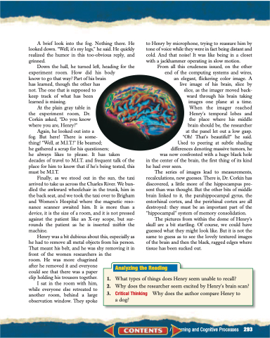
From all this crudeness issued, on the other end of the computing systems and wires, an elegant, flickering color image. A live image of his brain, slice by slice, as the imager moved back- ward through his brain taking images one plane at a time. When the imager reached Henry’s temporal lobes and the place where his middle brain should be, the researcher at the panel let out a low gasp. “Oh! That’s beautiful!” he said. Used to peering at subtle shading differences denoting massive tumors, he was now confronted with a huge black hole in the center of the brain, the first thing of its kind
he had ever seen.
The series of images lead to measurements,
recalculations, new guesses. There is, Dr. Corkin has discovered, a little more of the hippocampus pre- sent than was thought. But the other bits of middle brain linked to it, the parahippocampal gyrus, the entorhinal cortex, and the perirhinal cortex are all destroyed: they must be an important part of the “hippocampal” system of memory consolidation.
The pictures from within the dome of Henry’s skull are a bit startling. Of course, we could have guessed what they might look like. But it is not the same to guess as to see the lovely textured images of the brain and then the black, ragged edges where tissue has been sucked out.
   I sat in the room with him, while everyone else retreated to another room, behind a large observation window. They spoke
1. What types of things does Henry seem unable to recall?
2. Why does the researcher seem excited by Henry’s brain scan?
Analyzing the Reading
3. Critical Thinking a dog?
Why does the author compare Henry to
 Unit 4 / Learning and Cognitive Processes 293


