
Page 25 - Sonoma County Gazette 3-19
P. 25
MORE on the Marvel that is the Eye
Every student of anatomy has heard it a thousand times: “Structure Follows Function.” We need only to study the structure of a particular organ to glean precious insight into its function. When looked at closely, the structure of the eye is nothing less than astounding. Before we can discuss the health or disease of the eye, we must consider its structure.
Let’s start with the globe itself: the “eyeball” is made of a tough connective tissue that retains its shape and size even when subjected to constant changes in pressure. This material makes up the white sclera of the globe. With every pulse, blood entering the eye causes changes in the pressure and internal volume of the eye, and yet, we do not perceive changes in our visual image. The eye is like a camera built out of a balloon, yet it still takes fantastic pictures.
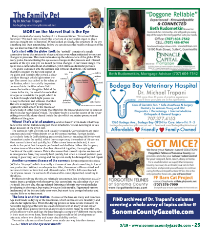
chamber occupies the forward aspect of
the globe and contains the cornea, a clear
window through which light enters the
eye. The cornea is attached to the sclera at
the limbus, which is crucially important
because this is the filter where fluid
leaves the inside of the globe. Behind the
cornea is the iris, the colorful muscle that
enlarges or constricts the pupil, which is
the hole through which light passes on
its way to the lens and vitreous chamber.
The lens is supported by suspensory
ligaments which attach to the muscular
ciliary body. It is the ciliary body that stretches the lens and allows us to focus on objects in our near field of vision. The ciliary body is also the source of the never- ending river of fluid pro-duced inside the eye which maintains pressure and inflation of the globe.
The globe is divided into the anterior and vitreous chambers. The anterior
iris lens cornea
sclera retina
choroid
Whew! That’s a lot of anatomy, and we haven’t even made it half way back to the retina! But knowing just these structures, we can now begin to talk about diseases of the eye it-self.
The cornea is right up front, so it is easily wounded. Corneal ulcers are quite common and occur when objects strike the corneal surface. Foreign bodies, particularly foxtails (self-planting grass seeds), have an amazing ability to work themselves under the eyelid, where they scrape away the surface of the cornea. Most corneal ulcers heal quickly, but if the cornea becomes infected, it may
erode to the point that the eye is perforated and de-flates. When this happens,
the structures of the anterior chamber often stick together, dis-rupting the function of the optic camera. This is the reason that corneal injuries are treat-ed
as emergencies. Sure, they usually heal quickly, but when a corneal problem goes wrong, it goes very, very wrong and the eye can easily be damaged beyond repair.
Another common disease of the cornea is keratoconjunctivitis sicca,
(KCS aka “dry eye”) which is actually a disease of tear glands resulting in loss of tear production. Without an adequate tear film, the cornea is ill-nourished and becomes irritated or infected, and often ulcerates. If KCS is left uncontrolled,
the dryness causes the cornea to thicken and be-come pigmented, resulting in blindness.
Problems involving the iris are relatively uncommon. Iris dysfunction usually results from a problem with the nerves that control iris muscle rather than the iris itself. Iris atro-phy, the age related thinning of the iris may result in holes developing in the organ, but typically causes little trouble. Pigmented tumors (melanoma) of the iris are fairly com-mon, but most have benign behavior and require little or no treatment.
anterior chamber
optic nerve ARTWORK BY HOLLY FISCHER
FIND archives of Dr. Trapani’s columns covering a whole array of topics online @
SonomaCountyGazette.com
The lens is another matter. Diseases of the lens are many and common. Age itself leads to drying of the lens tissue, which decreases lens flexibility and leads to far-sightedness. When the drying process is more severe it creates the noticeable fogging of the lens (nu-clear sclerosis) seen in many of our senior
dogs. High blood glucose levels in diabetes allow excess sugar to enter the lens, which attracts water and fogs the lens through a completely different mechanism. In their most extreme form, these lens changes result in the development of cataracts, where lens clarity and some visual ability are lost.
Two entire columns and we haven’t even made our way into the vitreous chamber! More on the eye next month!
3/19 - www.sonomacountygazette.com - 25
