
Page 42 - NMHBASummer2019
P. 42
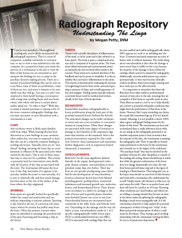
                                  If you’ve ever attended a Thoroughbred yearling sale, you’ve likely encountered the radiograph repository. This is a room of
computers, available exclusively to veterinar- ians, to use to view x-rays submitted to the sale company. As a prospective buyer you retain the services of a veterinarian at the sale to view the films of the horses you are interested in, and interpret the findings for you to assist in the purchase decision making process. There are a myriad of potential findings that can be seen on x-ray films; some of them are problematic, some of them are not, and some it remains to be seen which way they will go. Last year at a sale I was surprised to find myself having a conversation with a long-time yearling buyer and race horse owner, who when told about a certain abnor- mality asked me, “So what is that!?” With that in mind it seemed pertinent to discuss a few of the more common radiographic findings that you may encounter in your discussions with a veterinarian at a sale.
CHIPS
Any racehorse owner is likely quite familiar
with bone chips. While hearing this has been discovered as a new findings in your racehorse often strikes fear, you may be surprised to learn that this can be a relatively common finding in yearling sale horses. Typically, these are an “inci- dental” finding, meaning the horse has no overt lameness or effusion of the associated joint when viewed by the buyer. This is one of those issues that may or may not be a problem. The severity or potential need for intervention rests chiefly with the location of the chip as associated with the joint. The veterinarian will assess the loca- tion of the chip, determine if it appears to be articular (within the joint) or non-articular, if it is new or relatively old, and how much damage has been done to the rest of the surface of the bone where the chip originated.
SPURRING
Bone spurs are typically looked for specifically within the carpal and tarsal joints and can indicate impending or present arthritis. Spurring is also found at the site of a previous soft tissue injury, where the tendon has pulled away from the bone. Often the primary concern when spurs are identified is assessing the potential risk of the spur fracturing and becoming a chip in the future.
TARSITIS
Tarsitis is the scientific description of inflammation of the tarsal, or hock, joints and is also referred to as bone spavin. The hock is quite a complicated struc- ture and is composed of 4 separate joints. The lower joints (distal intertarsal and tarsometatarsal joints) are primarily the sites involved when tarsitis is dis- covered. These joints serve as shock absorbers of the hindlimb and can be prone to instability. It is this in- stability that can lead to inflammation in the joints. The severity is determined by evaluating the amount of roughening found in these joints, associated spur- ring or presence of chips, and overall appearance of the joint margins. Finding tarsitis typically indicates a potential future need for medical intervention, usually in the form of hock injections.
SESAMOIDITIS
Sesamoiditis manifests radiographically as
boney proliferation along the back part of the proximal sesamoid bones (behind the fetlock). The associated changes can be mild, moderate, or severe and can occur secondary to concussive trauma in young horses. Often these changes
are associated with some degree of irritation or damage to the branches of the suspensory liga- ment that attaches on the sesamoid. Rest is the primary intervention required for this condi- tion, however closer examination and sometimes further diagnostics, such as suspensory branch ultrasound, is warranted.
OSTEOCHONDRITIS
Believed to be the most significant skeletal disorder in the equine developmental ortho- pedic disease complex, osteochondritis is more prevalent today than ever before. There has
been no one specific predisposing cause identi- fied for the development of osteochondritis, however numerous factors have been blamed including genetic predisposition, nutritional imbalances and increased growth rate, endocrine factors, and biomechanical forces. These lesions occur secondary to a defect in cartilage at the ends of bones and in growth plates; therefore, osteochondral lesions can occur in any joint. Osteochondral lesions are encountered most commonly in the stifle, hock, and fetlocks, how- ever. Depending on the damage and the way the defect occurs, osteochondritis results in 1 of 2 specific radiographically visible lesion types: OCD or subchondral bwwewone cyst (SBC). OCD is associated with cartilage flaps that can
become ossified and visible radiographically where SBCs appears as a void or an infolding into the bone. Common associated symptoms are joint ef- fusion with or without lameness. The tricky thing about osteochondritis is that often the damage is more severe than is visible on the x-ray, because the majority of the damage is done within the cartilage which cannot be assessed by radiography. Additionally, osteochondral lesions may resolve spontaneously, or they may become clinically evident problems when horses begin training and have strain put on the affected joints.
It is imperative to remember that these sale
films have been taken within a predetermined amount of time prior to the sale, meaning they are
a snapshot of that horse at a specific point in time. These films are used as a tool to try to help identify any current or potential orthopedic conditions that might limit the performance of that horse in the future. However, like I enjoy reminding my clients, the crystal ball sometimes fogs up if it’s too humid outside. Meaning, it is not possible to know 100% what a given radiograph finding means for the future of that horse. We veterinarians use facts and accumulated data to make inferences about what we are seeing on the radiographs presented to us. Another important point to bear in mind is that
the majority of the time, the veterinarian is assessing the radiographs only. There is typically no formal exam performed on the horse by the veterinarian, and certainly not to the degree of the traditional “Pre-purchase Exam” that may be involved in the purchase of horses for other disciplines or outside of the yearling sale setting. Buyers should keep in mind that while the general conformation of the horse does not change dramatically over time, the way their bones and soft tissue respond to the rigors of training is a fluid situation. That being said, you as the buyer must decide on your level of risk tolerance and work with a veterinarian who understands this as well. Simply stated, risk tolerance is what you can and cannot live with. There is no perfect horse and there will never be a perfect set of x-rays. Knowing what conditions are a deal breaker, and which you are willing to manage should they become an issue in the future makes deciphering the radiographic findings a much more manageable task. It is the veterinarians objective to help explain the potential consequence of what is being seen “today” in the films and provide interpretation of what it could mean for the future. Thus, having a good working relationship with the veterinarian viewing the films for you is of paramount importance.
Radiograph Repository
Understanding The Lingo
by Megan Petty, DVM
 40 New Mexico Horse Breeder
