
Page 95 - Speedhorse October 2018
P. 95
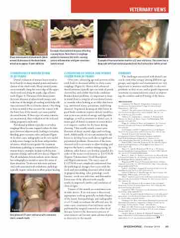
Figure 3
Sharp enamel points of cheek teeth (black arrows) & abrasions to the cheek (white arrow) on an approx. 11-year-old horse.
Conditions of Mature Horse (5-20 years)
Dental evaluation of mature horses tends to focus heavily on sharp enamel points and maloc- clusion of the cheek teeth. Sharp enamel points occur normally along the outer edge of the upper cheek teeth and along the inside edge of lower teeth (Figure 3). Flotation of the sharp points decreases abrasion of adjacent soft tissues, and reduction of the height of overlong teeth helps alle- viate unwanted effects of malocclusion. One thing to keep in mind is that excessive bit contact with the lower bars of the mouth can cause painful, ulcerated lesions. If these type of contact injuries are encountered, then evaluation of the tack and riding program is indicated.
Periodontal problems typically occur when roughage fibers become trapped within the slim space between adjacent teeth, leading to irritation, bleeding, gum recession, odor, and pain (Figure
4). In these cases, radiography can be very useful to help assess changes in the bone and periodon- tal tissues, which in turn guides the treatment. Endodontic pathology is commonly identified in mature horses: examples include tooth fracture, exposure of pulp, and tooth root abscess (Figure 5). Not all endodontic lesions indicate active disease, but radiography is needed to assess the status of these situations. Tooth root infections and non- vital teeth associated with endodontic pathology typically require extraction to allow proper healing.
Figure 6
Oral photo of 27-year-old horse with numerous short, smooth teeth.
Figure 5
Example of fractured molar tooth in a 12-year-old horse. The crown has a deep split with feed material packed into the fracture line (white arrow).
Figure 4
Example of periodontal disease affecting a young horse. Note there is roughage packed in-between the teeth, causing severe inflammation and gum recession (white arrow).
Conditions of senior age Horses (older tHan 20 years)
Over time, advancing age and attrition of the teeth leads to decreased ability to chew coarse roughage (Figure 6). Horses with advanced dental attrition typically spit-out wads of poorly chewed hay and exhibit thin body condition. Besides dental problems, it’s important to keep in mind there’s a long list of non-dental factors to consider when looking at an older thin horse (e.g. nutritional status, parasitism, underlying diseases). In general, keeping an older horse in good body condition requires dietary modifica- tion to increase intake of energy and digestible roughage, as well as attention to dental care. A major goal of dental treatment in senior horses
is to improve comfort for the horse during chewing. This usually entails conservative flotation of sharp enamel edges and overlong teeth. Additionally, it’s not uncommon for old horses to develop loose teeth due to significant periodontal problems. Extraction of the loose, diseased teeth is necessary to allow healing and improve the horse’s comfort during eating. In addition, older horses can develop a painful dis- order of the incisor teeth known as ‘E.O.T.R.H’ (Equine Odontoclastic Tooth Resorption
and Hypercementosis). The exact cause of E.O.T.R.H is not completely understood, but the pathological changes that occur with the teeth and surrounding structures often result in gingival bleeding, odor, pathologic tooth fracture, tooth root infection, and discomfort. Extraction of the affected teeth usually
results in improved comfort and resolution of clinical signs.
Tumors of the mouth are sometimes seen in older horses. If an oral mass is discovered, appropriate work-up generally includes biopsy of the lesion, histopathology, and radiography or a CT study to evaluate the affected area. In some cases, treatment of an oral mass can be performed by surgical removal or other local therapy, however the destructive process of some tumors can become quite advanced prior to discovery in the mouth.
suMMary
The challenges associated with dental care can be somewhat unique among different age groups, and regular oral examinations are very important to monitor oral health, to discover problems as they occur, and to guide important treatment recommendations aimed at improv- ing the comfort and well-being of the horse.
BiBliography
Committee AN, Board A. Nomenclatue Committee of the American Veterinary Dental College.Veterinary Dental Nomenclature. 2007:1-12.
Griffin C. The gold standard of dental care: the juvenile horse. Vet Clin North Am Equine Pract. 2013;29(2):487-504, vii - viii. doi:10.1016/j.cveq.2013.04.004.
Tulleners, E., Schumacher, J., Johnston, J., Richardson D. Pharynx. In: Auer J, ed. Equine Surgery. Vol Philadelphia: W.B. Saunders; 1992:453-459.
Wiggs, R., Lobprise H. Basics of Orthodontics. In: Veterinary Dentistry, Principles and Practice1. Vol Philadelphia: Lippencot; 1997:438-441.
Earl Gaughan and RD. Congenital Diseases of the Equine Head. In: Vetterinary Clinics of North America. Vol ; 1993:93-110.
Swerczek T, Lieto L CE. Scientific Papers
Developmental Defects of Enamel in American Saddlebred Foals With Epitheliogenesis Imperfecta. J Equine Vet Sci. 2004;24(September):386-390. doi:10.1016/j.jevs.2004.08.008.
Ramzan PHL, Dixon PM, Kempson SA, Rossdale PD. Dental dysplasia and oligodontia in a Thoroughbred colt. Equine Vet J. 2001;33(1):99-104.
Morse CC, Saik JE, Richardson DW, Fetter a. W. Equine Juvenile Mandibular Ossifying Fibroma. Vet Pathol. 1988;25(6):415- 421. doi:10.1177/030098588802500603.
Brown, C., Baker, D., Barker I. Oral Cavity. In: Maxie M, Jubb K, Kennedy P, Palmer N, eds. Pathology of Domestic Animals. Vol 5th ed. Edinburgh: Elsevier Saunders; 2007:3-32.
Waguespack RW, Taintor J. Paranasal sinus disease in horses. Compend Contin Educ Vet. 2011;33(2):E1-E12. http://www.ncbi.nlm. nih.gov/pubmed/21870345.
Lamb C, Schelling S. Congenitial aneurysmal bone cyst in the mandible of a foal. Eq Vet J. 1989;21(2):130-132.
Nickels F, Tulleners E. Nasal Passages. In: Equine Surgery. Vol Philadelphia: WB Saunders; 1992:433-446.
Allen T. Incidence and severity of abrasions of the buccal mucosa adjacent to the cheek teeth in 199 horses. In: American Assoc Eq Prac. Vol ; 2004:31-36.
Tremaine H. Advances in the treatment of diseased equine cheek teeth. Vet Clin North Am Equine Pract. 2013;29(2):441-465. doi:10.1016/j.cveq.2013.04.013.
Carmalt J.L. WD. Treatment of a valve diastema in two horses. Eq Vet Educ. 2004;16(4):188-192.
Foster DL. The gold standard of dental care for the adult perfor- mance horse. Vet Clin North Am Equine Pract. 2013;29(2):505-519, viii. doi:10.1016/j.cveq.2013.04.012.
Niederman CN. How to Incorporate Nutritional Discussions and Nutritional Alterations As Components of Equine Dental Care. In: Proceedings, Am Assoc Eq Prac. Vol 60. ; 2014:490-493.
Baker, G.J. CKJ. Dentistry in the Geriatric Horse. In: Bertone J, ed. Equine Geriatric Medicine and Surgery. Vol I. St. Louis: Saunders Elsevier; 2003:51-58.
Staszyk C, Bienert A, Kreutzer R, Wohlsein P, Simhofer H. Equine odontoclastic tooth resorption and hypercementosis. Vet J.
2008;178(3):372-379. doi:10.1016/j.tvjl.2008.09.017.
SPEEDHORSE, October 2018 93
veterinary views
