
Page 14 - HOME TEST OB SONO 260
P. 14
SONO 260 2016 Prof. Ana Zayas Ed.D(s)
Anatomy and physiology
54) A patient with a positive pregnancy test is referred to the ultrasound department with a history of painless vaginal bleeding. Sonographically, you visualize a single live intrauterine gestation. The crown rump length is consistent with an 8 week pregnancy. Within the fetal skull, you visualize a round, anechoic structure. What does this most likely represent?
a. Megacisternamagna
b. Cystic hygroma
c. Rhombencephalon
d. Thickened nuchal translucency
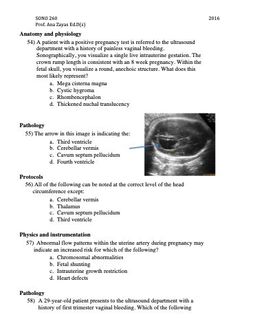
Pathology
55) The arrow in this image is indicating the:
a. Thirdventricle
b. Cerebellar vermis
c. Cavumseptumpellucidum d. Fourth ventricle
Protocols
56) All of the following can be noted at the correct level of the head circumference except:
a. Cerebellarvermis
b. Thalamus
c. Cavumseptumpellucidum d. Third ventricle
Physics and instrumentation
57) Abnormal flow patterns within the uterine artery during pregnancy may indicate an increased risk for which of the following?
a. Chromosomalabnormalities b. Fetal shunting
c. Intrauterinegrowthrestriction d. Heart defects
Pathology
58) A 29-year-old patient presents to the ultrasound department with a history of first trimester vaginal bleeding. Which of the following
