
Page 158 - Libro 2
P. 158
138 PART 3 — PERIPHERAL ARTERIAL
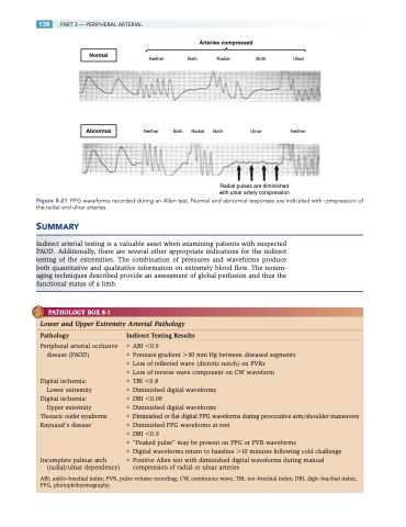
Normal
Abnormal
Neither
Neither
Both Radial Both
Both Radial Both Ulnar
Radial pulses are diminished with ulnar artery compression
Ulnar
Neither
Arteries compressed
Figure 8-21 PPG waveforms recorded during an Allen test. Normal and abnormal responses are indicated with compression of the radial and ulnar arteries.
SUMMARY
Indirect arterial testing is a valuable asset when examining patients with suspected PAOD. Additionally, there are several other appropriate indications for the indirect testing of the extremities. The combination of pressures and waveforms produce both quantitative and qualitative information on extremity blood flow. The nonim- aging techniques described provide an assessment of global perfusion and thus the functional status of a limb.
PATHOLOGY BOX 8-1
Lower and Upper Extremity Arterial Pathology
Pathology Indirect Testing Results
Peripheral arterial occlusive disease (PAOD)
Digital ischemia: Lower extremity
Digital ischemia: Upper extremity
Thoracic outlet syndrome Raynaud’s disease
Incomplete palmar arch (radial/ulnar dependency)
• ABI 0.9
• Pressure gradient 30 mm Hg between diseased segments
• Loss of reflected wave (dicrotic notch) on PVRs
• Loss of reverse wave component on CW waveform
• TBI 0.8
• Diminished digital waveforms
• DBI 0.09
• Diminished digital waveforms
• Diminished or flat digital PPG waveforms during provocative arm/shoulder maneuvers
• Diminished PPG waveforms at rest
• DBI 0.9
• “Peaked pulse” may be present on PPG or PVR waveforms
• Digital waveforms return to baseline 10 minutes following cold challenge
• Positive Allen test with diminished digital waveforms during manual compression of radial or ulnar arteries
ABI, ankle–brachial index; PVR, pulse volume recording; CW, continuous wave; TBI, toe–brachial index; DBI, digit–brachial index; PPG, photoplethysmography.
