
Page 176 - Libro 2
P. 176
156
PART 3 — PERIPHERAL ARTERIAL
Right common carotid artery Right vertebral artery
Subclavian artery Axillary artery
Brachiocephalic trunk
arteries are the first major branches of both the right and left subclavian arteries. The left vertebral artery may originate directly from the aortic arch in 4% to 6% of patients.1 Also arising from the subclavian arteries are the thyrocervical and costocervical trunks. These arteries can be distinguished from the vertebral arteries by their multiple branches and lower end-diastolic flow velocities.
The subclavian arteries exit the chest through the thoracic outlet (Fig. 10-2). In the course of the subcla- vian artery, there are three spaces or distinct sites for potential compression. The subclavian artery passes over the first rib between the anterior and middle scalene muscles through the scalene triangle. The subclavian vein passes superficial to the anterior sca- lene muscle and bypasses the scalene triangle. The costoclavicular space is bound by the clavicle and first rib and is the next area of possible compression. It is traversed by all three components of the neuro- vascular bundle. The third (most lateral) space is the pectoralis minor space. It is infrequently involved in symptomatic compression.2 Upper extremity arterial symptoms may be caused by impingement within the thoracic outlet. The effects on the subclavian artery may include stenosis, aneurysmal dilatation, laminar thrombus, and dynamic compression with arm abduction. The subclavian artery is renamed the axillary artery at the lateral margin of the first rib. The axillary artery lies deep to the pectoralis major and minor muscles. Within the axilla, it can be found deep to the axillary fat pad.
The axillary artery transitions to the brachial ar- tery at the level of the inferolateral border of the teres major muscle. This muscle cannot be routinely iden- tified by duplex ultrasound. Here, the artery takes a more superficial course in the medial arm between
Profunda brachial artery
Brachial artery
Radial artery
Aortic arch
Ulnar artery Interosseous artery
Superficial palmar arch Deep palmar arch
Figure 10-1
ity arteries.
Schematic drawing of the principle upper extrem-
The subclavian arteries originate in the chest, usu- ally arising from the brachiocephalic artery on the right and directly from the aortic arch on the left. The brachiocephalic artery (also known as the innominate artery) is the first major branch of the aortic arch and divides into the right common carotid artery and the right subclavian artery. Rarely, the right subclavian artery may originate directly from the aorta distal to the left subclavian artery in what is known as a retro- esophageal subclavian artery, or an aberrant subcla- vian artery. An aberrant right subclavian artery often arises from a dilated segment of the proximal de- scending aorta known as a Kommerell’s diverticulum. Although most patients are asymptomatic, some may have difficulty with swallowing from compression of the esophagus (dysphagia lusoria) by the abnormally positioned subclavian artery. Palsy of the recurrent laryngeal nerve can occur with this anatomic varia- tion and is called Ortner’s syndrome.
The left subclavian artery takes its origin directly from the aortic arch as the third major branch fol- lowing the left common carotid artery. The vertebral
Brachial plexus Clavicle (cut)
Subclavian artey
Subclavian vein
First rib (cut)
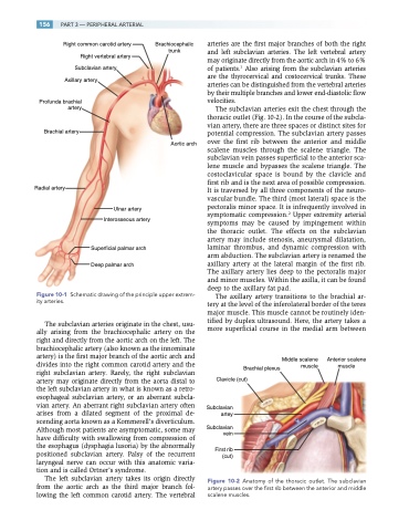
Middle scalene muscle
Anterior scalene muscle
Figure 10-2 Anatomy of the thoracic outlet. The subclavian artery passes over the first rib between the anterior and middle scalene muscles.
