
Page 355 - Libro 2
P. 355
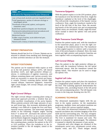
    TABLE 22-1
Indications for Hepatoportal Duplex Ultrasound
Liver cirrhosis both alcoholic and viral, hepatitis B and C Portal hypertension, ascites of unknown etiology or
esophageal varices
Thrombosis of the portal, splenic, and superior
mesenteric veins
Budd-Chiari syndrome (hepatic vein thrombosis) Preinterventional/postinterventional procedures and
monitoring of portosystemic shunts
Abdominal trauma
Sudden onset of ascites, acute abdominal pain, elevated D-dimer
Patients with a history of abdominal malignancy
PATIENT PREPARATION
Patients should fast for 8 to 12 hours. Patients are in- structed to abstain from smoking and chewing gum as these activities introduce air into the stomach.
PATIENT POSITIONING
The patient can be examined while supine as well as in the left posterior oblique (LPO), left lateral decubitus (LLD), and right posterior oblique po- sitions. A combination of sagittal, transverse, and oblique scanning planes and various acoustic win- dows are necessary to assess the portal and hepat- ic vessels. Primary scanning planes include right coronal oblique, transverse epigastric, transverse right costal margin, left coronal oblique, and sagit- tal left lobe.
Right Coronal Oblique
The right coronal oblique scanning plane using an intercostal approach provides a window that pro- vides excellent visualization of the porta hepatis. When patients present with large amounts of asci- tes and bowel gas, this increases abdominal girth, making it very difficult to image the liver and hepa- toportal venous anatomy. The intercostal approach results in the following: Doppler angles varying from 0° to about 60°, decreases the anterior poste- rior distance, uses the liver as an acoustic window, and facilitates diagnostic imaging when patients are not compliant with holding their breath. To obtain this scanning plane, position the patient in the LPO or LLD positions. Place the transducer in the in- terspace parallel to the rib margins, aimed at the hepatic hilum.
22 — The Hepatoportal System 335 Transverse Epigastric
With the patient supine or in the LPO position, place the transducer over the left lobe of the liver. Angle the transducer cephalad at the level of the diaphragm. This will provide visualization of the hepatic venous confluence. Next, angle the transducer caudad at the level of the left lobe of the liver. Here, the ascend- ing branch of the left portal vein and accompanying hepatic artery are visualized. Finally, angle the trans- ducer caudad to detect the splenic vein and portal confluence.
Right Transverse Costal Margin
Position the patient supine and, with the transducer in the transverse plane, place it over the right cos- tal margin at the midclavicular line. The transducer is then angled superior to inferior until a transverse view of the porta hepatis is obtained. Here, the main portal and right anterior and posterior branches are seen together with accompanying hepatic artery.
Left Coronal Oblique
Place the patient in the right posterior oblique po- sition with the transducer between the left lateral intercostal spaces. Aim the transducer toward the splenic hilum. This window can be used to image the splenic vein.
Sagittal Left Lobe
Position the patient supine and place the transducer in a longitudinal plane midline over the left lobe of the liver. This scanning approach demonstrates the left hepatic vein, ascending branch of the left portal vein, and accompanying artery. The left hepatic vein is seen in the long axis draining into the IVC.
SCANNING TECHNIQUE
The ultrasound examination begins with the pa- tient in the supine position. As the examination progresses, multiple views and patient positions are used to obtain the various required images.
Very often, the ultrasound evaluation of the hepa- toportal circulation also includes imaging of the liver itself and the surrounding area. The size, echogeni- city, and surface contour of the liver are documented. The liver is examined for masses and cysts. The spleen is evaluated for splenomegaly. The abdomen is scanned to identify the presence of any ascites.
Grayscale and color images are obtained of the extrahepatic portal vein, intrahepatic portal veins, hepatic veins, and IVC. These images should
