
Page 51 - Libro 2
P. 51
3 — Venous Physiology
31
Ppv
“Pcv” Pcv
Rv
Qv
Obstructed main venous channel
Capillary bed
Collateral veins
Peripheral veins
Diaphragm
Abdomen Thorax
Closed containers
Right atrium
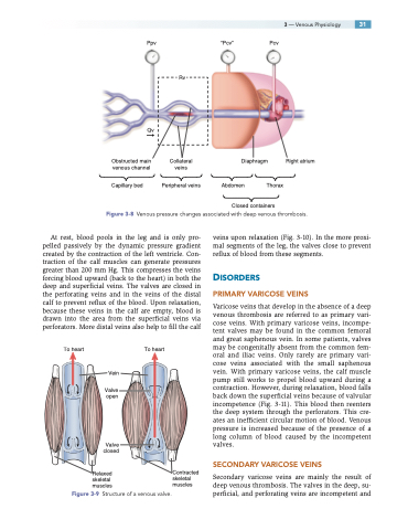
Figure 3-8 Venous pressure changes associated with deep venous thrombosis.
At rest, blood pools in the leg and is only pro- pelled passively by the dynamic pressure gradient created by the contraction of the left ventricle. Con- traction of the calf muscles can generate pressures greater than 200 mm Hg. This compresses the veins forcing blood upward (back to the heart) in both the deep and superficial veins. The valves are closed in the perforating veins and in the veins of the distal calf to prevent reflux of the blood. Upon relaxation, because these veins in the calf are empty, blood is drawn into the area from the superficial veins via perforators. More distal veins also help to fill the calf
veins upon relaxation (Fig. 3-10). In the more proxi- mal segments of the leg, the valves close to prevent reflux of blood from these segments.
DISORDERS
PRIMARY VARICOSE VEINS
Varicose veins that develop in the absence of a deep venous thrombosis are referred to as primary vari- cose veins. With primary varicose veins, incompe- tent valves may be found in the common femoral and great saphenous vein. In some patients, valves may be congenitally absent from the common fem- oral and iliac veins. Only rarely are primary vari- cose veins associated with the small saphenous vein. With primary varicose veins, the calf muscle pump still works to propel blood upward during a contraction. However, during relaxation, blood falls back down the superficial veins because of valvular incompetence (Fig. 3-11). This blood then reenters the deep system through the perforators. This cre- ates an inefficient circular motion of blood. Venous pressure is increased because of the presence of a long column of blood caused by the incompetent valves.
SECONDARY VARICOSE VEINS
Secondary varicose veins are mainly the result of deep venous thrombosis. The valves in the deep, su- perficial, and perforating veins are incompetent and
To heart
To heart
Vein
Valve open
Valve closed
Relaxed skeletal muscles
Contracted skeletal muscles
Figure 3-9 Structure of a venous valve.
