
Page 80 - Libro 2
P. 80
60
PART 2 — CEREBROVASCULAR
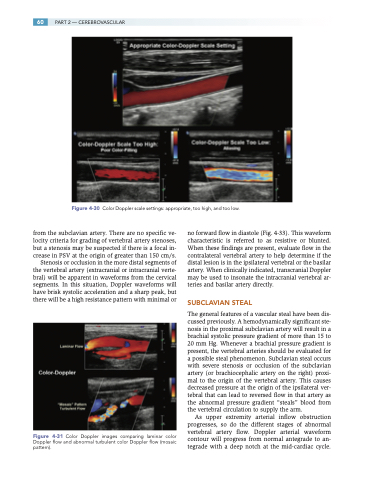
Figure 4-30 Color Doppler scale settings: appropriate, too high, and too low.
from the subclavian artery. There are no specific ve- locity criteria for grading of vertebral artery stenoses, but a stenosis may be suspected if there is a focal in- crease in PSV at the origin of greater than 150 cm/s.
Stenosis or occlusion in the more distal segments of the vertebral artery (extracranial or intracranial verte- bral) will be apparent in waveforms from the cervical segments. In this situation, Doppler waveforms will have brisk systolic acceleration and a sharp peak, but there will be a high resistance pattern with minimal or
Figure 4-31 Color Doppler images comparing laminar color Doppler flow and abnormal turbulent color Doppler flow (mosaic pattern).
no forward flow in diastole (Fig. 4-33). This waveform characteristic is referred to as resistive or blunted. When these findings are present, evaluate flow in the contralateral vertebral artery to help determine if the distal lesion is in the ipsilateral vertebral or the basilar artery. When clinically indicated, transcranial Doppler may be used to insonate the intracranial vertebral ar- teries and basilar artery directly.
SUBCLAVIAN STEAL
The general features of a vascular steal have been dis- cussed previously. A hemodynamically significant ste- nosis in the proximal subclavian artery will result in a brachial systolic pressure gradient of more than 15 to 20 mm Hg. Whenever a brachial pressure gradient is present, the vertebral arteries should be evaluated for a possible steal phenomenon. Subclavian steal occurs with severe stenosis or occlusion of the subclavian artery (or brachiocephalic artery on the right) proxi- mal to the origin of the vertebral artery. This causes decreased pressure at the origin of the ipsilateral ver- tebral that can lead to reversed flow in that artery as the abnormal pressure gradient “steals” blood from the vertebral circulation to supply the arm.
As upper extremity arterial inflow obstruction progresses, so do the different stages of abnormal vertebral artery flow. Doppler arterial waveform contour will progress from normal antegrade to an- tegrade with a deep notch at the mid-cardiac cycle.
