
Page 11 - Libro vascular I
P. 11
Chap-01.qxd 29~8~04 13:19 Page 2
2
PERIPHERAL VASCULAR ULTRASOUND
therapeutic procedures, such as angioplasty, rather than diagnostic angiograms.
Vascular ultrasound examinations rely on the use of ultrasound to produce a black and white anatom- ical image that can demonstrate the presence of disease along an arterial wall or the presence of thrombus in a vein. Doppler ultrasound can provide a functional map in the form of a color flow image, which displays the blood flow in arteries and veins. Spectral Doppler analysis enables Doppler wave- forms to be recorded from vessels. It is then possi- ble to visualize changes in flow patterns in vessels and calculate velocity measurements, enabling the sonographer to grade the severity of the vascular disease (Fig. 1.1).
Arterial disease is one of the major causes of mor- bidity and mortality in the developed world. There are many risk factors associated with the develop- ment of arterial disease, but it is widely accepted that tobacco smoking is one of the primary causes. Atherosclerotic plaques develop over time, leading to arterial obstruction or embolization. Radiologists and surgeons are able to perform a variety of proce- dures to treat arterial disorders. Angioplasty involves the use of a balloon mounted on the end of a
An example of a carotid ultrasound scan showing how B-mode imaging, color flow imaging and spectral Doppler are used to investigate a stenosis.
catheter which is guided, using angiography, to the area of stenosis (narrowing) or occlusion (blockage). The balloon is then positioned across the stenosis or occlusion and inflated for a short period of time, to
Figure 1.1
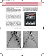
AB
Figure 1.2 A: An angiogram demonstrating a significant stenosis in the right common iliac artery (arrow). B: The stenosis has been dilated by percutaneous balloon angioplasty.
