
Page 152 - Libro vascular I
P. 152
Chap-10.qxd 29~8~04 14:48 Page 143
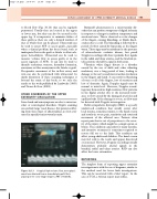
to blood flow (Fig. 10.13) that can be regularly punctured. Usually they are located in the upper or lower arm, but they can also be created in the upper leg. It is imperative to minimize failure of these grafts as there are only a limited number of sites at which they can be placed. Ultrasound can be used to assess AVF or access grafts, especially when a clinical problem has been found, such as inadequate flow in the graft or fistula to allow ade- quate hemodialysis. Ultrasound can be used to measure volume flow in access grafts or in the access segment of AVFs. It can also be used to identify occlusions, stenoses, thrombus formation, aneurysms or false aneurysms in the fistula or graft. Preoperative assessment of the in-flow artery and vein can also be performed with ultrasound. In depth discussion of these scanning techniques is beyond the remit of this book, so we refer the reader to the work published by Landwehr (1995) and Deane & Goss (2001).
OTHER DISORDERS OF THE UPPER
EXTREMITY CIRCULATION
Some hand and arm symptoms are due to microvas- cular or neurological disorders. Duplex scanning can exclude large vessel disease, but patients suffer- ing from these types of abnormalities are best eval- uated in specialist microvascular units.
Figure 10.13 A typical high-volume flow, monophasic waveform obtained from a hemodialysis graft. Note aliasing in the color image despite a PRF of 7000 Hz.
DUPLEX ASSESSMENT OF UPPER EXTREMITY ARTERIAL DISEASE
143
Raynaud’s phenomenon is a microvascular dis- order that can produce symptoms of digital ischemia in response to changes in ambient temperature and emotional state. This is observed as color changes of the fingers, causing blanching, or bluish discol- oration due to cold. The blanching is followed by a period of rubor caused by hyperemia as the fingers warm. These signs may be mistaken for the presence of atherosclerotic occlusive disease, but pencil Doppler recordings will detect pulsatile flow signals in the radial and ulnar arteries, and the brachial sys- tolic pressure should be equal in both arms.
Vibration white finger disease is a disorder caused by the use of drills and other vibrating machinery over a long period of time, leading to damage to the nerves and microvascular circulation in the fingers and hand. It can result in blanching of some or all of the fingers, loss of sensation and loss of dexterity. Again, Doppler signals may be normal to wrist level. However, Doppler record- ings may demonstrate high-resistance flow patterns in the digital arteries due to the increased resist- ance to flow caused by the damaged arterioles and capillary beds. If the damage is severe, no flow may be detected with Doppler interrogation.
Reflex sympathetic dystrophy (RSD) is a poorly understood condition that usually occurs after local trauma, sometimes minor, to the hand or arm and results in severe pain, sensitivity and restricted movement of the affected area. Patients often report pain that is out of proportion to the sever- ity of the injury, which might be a simple sprain or bruise. The condition can persist for many months, and intensive treatment is sometimes required to restore full use to the limb. This condition can affect young adults and children. The hand or arm may feel cold to the touch and appear discolored or cyanosed. However, Doppler recordings usually demonstrate pulsatile arterial signals in the brachial, radial and ulnar arteries. RSD can also affect the lower extremities.
REPORTING
The simplest form of reporting upper extremity investigations is with the use of diagrams, similar to the method used for lower limb investigations. This can be associated with a brief report. In the case of TOS, a written report may suffice.
