
Page 14 - DREAM 2047_English June 2021
P. 14
HEALTH
TV Venkateswaran
The SARS-CoV-2 virus in the droplets that we inhale finds a welcome home in the lining of the nose. The surface of the cells that form the lining of the respiratory tract, called epithelial cells, are rich in the cell-surface receptors, a type of proteins called angiotensin-converting enzyme 2 (ACE2). Typically, this receptor which helps regulate blood pressure, binds with a part of the viral spike protein called the receptor-binding domain (RBD). The virus, then, takes a foothold grabbing onto the ACE2 receptor, and enters the cells. Once inside, the virus hijacks the cell's machinery, reproducing myriad copies of itself. The viral copies emerge from the infected cell and invade new cells.
As the virus multiplies and more and more cells are invaded by the virus, the immune system works in overdrive. If not controlled in the initial stages, the virus marches into the alveoli of the lungs.
make it harder, at times even impossible, for you to breathe. As fluid collects inside the lungs, they carry less oxygen to your blood. That means your blood may not supply your organs with enough oxygen to survive. The patient would gasp for breath. If the oxygen level steadily falls down, the patient might suffer hypoxia.
Gas exchange in lungs
Like a tree diverge out from trunk, branch and twig, the lungs also branch out into bronchi, bronchioles, and air sacs called alveoli at the terminal end of bronchioles. The bifurcating tree structure of the lungs is so extensive that together the lungs
Through the mechanism of diffusion, O2 and CO2 are exchanged between the air and the pulmonary capillaries in alveoli.
contain approximately 2,400 kilometre of airways, about the distance of Chennai from Delhi. The total surface area of 300 to 500 million alveoli in the lungs is about 100 square meter, roughly the size of a tennis court. The walls of these alveoli are just one cell in thick. They are crisscrossed with tiny pulmonary capillaries blood vessels.
Oxygen circulation
Haemoglobin in the blood and myoglobin in the muscles are oxygen-carrying proteins. These proteins have an iron- containing group called ‛hemeʼ. Typically, there is about 12 to 20 gram of haemoglobin in every 100 ml of blood. The Fe2+ in the haemoglobin can reversibly bind with O2. Oxygen is a nonpolar molecule in which, due to the distributional arrangement of electrons, induces dipole. Fe2+ exerts a week ion-induced dipole attraction and binds to O2. Once in the blood, the oxygen is bound to haemoglobin in red blood cells, taken through the pulmonary vein to the heart, pumped into the systemic vascular system and, finally, brought to all the body cells. Oxygen absorbed by the haemoglobin in lungs is transported by blood to tissue
Can one rely on the smartphone SpO2 sensors? The simple answer is No. For computing the SpO2 levels, the beam of probe light has specific wavelengths of red and infrared. However, the smartphone cameras have regular white or yellowish flashlights. Therefore, they may not give adequately reliable readings.
14 dream2047/june2021
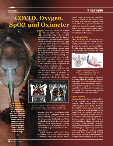
In the scarred lungs, on the right, there are much larger areas of darkness, representing parts of the lungs that are having difficulty transporting oxygen into the blood stream (Image: Oxford University)
Alveoli are lined with a single layer of cells, also rich in ACE2 receptors. Once these cells are infected, alveoli are destroyed, and the virus can enter the bloodstream. White blood cells react to the virus and infected cells and release inflammatory molecules called chemokines. More immune cells are summoned to target and destroy the virus- infected cells.
Alveoli became stuffed with fluid, white blood cells, mucus, and the detritus of destroyed lung cells. Clogged alveoli
