
Page 114 - Simplicity is Key in CRT
P. 114
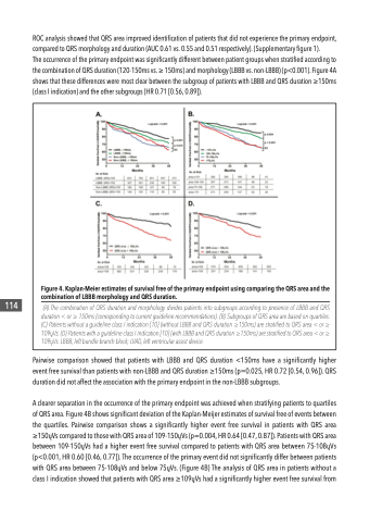
  Figure 4. Kaplan-Meier estimates of survival free of the primary endpoint using comparing the QRS area and the combination of LBBB morphology and QRS duration.
(A) The combination of QRS duration and morphology divides patients into subgroups according to presence of LBBB and QRS duration < or ≥ 150ms (corresponding to current guideline recommendations). (B) Subgroups of QRS area are based on quartiles. (C) Patients without a guideline class I indication [10] (without LBBB and QRS duration ≥150ms) are stratified to QRS area < or ≥ 109ųVs. (D) Patients with a guideline class I indication [10] (with LBBB and QRS duration ≥150ms) are stratified to QRS area < or ≥ 109ųVs. LBBB, left bundle branch block; LVAD, left ventricular assist device.
 114
 ROC analysis showed that QRS area improved identification of patients that did not experience the primary endpoint, compared to QRS morphology and duration (AUC 0.61 vs. 0.55 and 0.51 respectively). (Supplementary figure 1).
The occurrence of the primary endpoint was significantly different between patient groups when stratified according to the combination of QRS duration (120-150ms vs. ≥ 150ms) and morphology (LBBB vs. non-LBBB) (p<0.001). Figure 4A shows that these differences were most clear between the subgroup of patients with LBBB and QRS duration ≥150ms (class I indication) and the other subgroups (HR 0.71 [0.56, 0.89]).
Pairwise comparison showed that patients with LBBB and QRS duration <150ms have a significantly higher event free survival than patients with non-LBBB and QRS duration ≥150ms (p=0.025, HR 0.72 [0.54, 0.96]). QRS duration did not affect the association with the primary endpoint in the non-LBBB subgroups.
A clearer separation in the occurrence of the primary endpoint was achieved when stratifying patients to quartiles of QRS area. Figure 4B shows significant deviation of the Kaplan-Meijer estimates of survival free of events between the quartiles. Pairwise comparison shows a significantly higher event free survival in patients with QRS area ≥150ųVs compared to those with QRS area of 109-150ųVs (p=0.004, HR 0.64 [0.47, 0.87]). Patients with QRS area between 109-150ųVs had a higher event free survival compared to patients with QRS area between 75-108ųVs (p<0.001, HR 0.60 [0.46, 0.77]). The occurrence of the primary event did not significantly differ between patients with QRS area between 75-108ųVs and below 75ųVs. (Figure 4B) The analysis of QRS area in patients without a class I indication showed that patients with QRS area ≥109ųVs had a significantly higher event free survival from
