
Page 59 - Simplicity is Key in CRT
P. 59
ECG acquisition and analysis.
Standard supine 12-lead ECGs (filter range, 0.15 to 100 Hz; AC filter, 60 Hz, 25 mm/sec speed, 1 mm/mV) were obtained at baseline and before discharge. Blinded to outcomes, one experienced reader interpreted QRS morphology based on morphological features of the five different definitions. LBBB definitions used were the ones recommended by the AHA/ACC/HRS [9], the ones suggested by the ESC in 2006 [10] and 2009 [11] textbooks and the 2013 ESC guidelines [6], and finally the ECG definition proposed by Strauss et al. [12]; the criteria are summarized in Table 1. Notching in the QRS complex and slurring were defined according to Almer et al. [13]. Notching was defined as a sudden change, within a slope (waveform), in direction ≥90°; slurring was defined as sudden change in the slope of a waveform with a change in direction 0°-90°. QRS duration was always automatically measured by using ECG machine of two different vendors (Schiller CARDIOVIT CS-200 Excellence, Doral, FL, USA and ELI 350, Mortara Instrument,Inc., Milwaukee, WI, USA). In order to qualify for LBBB according to a specific definition, the ECG should comply with all required criteria for that definition.
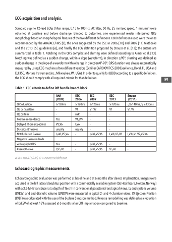
Table 1. ECG criteria to define left bundle branch block.
AHA (2009)
ESC 2006
ESC 2009
ESC 2013
Strauss (2011)
QRS duration
≥120ms
≥120ms
≥120ms
≥120ms
♂≥140ms, ♀≥130ms
QS or rS pattern
-
V1
V1,V2
V1
V1,V2
QS pattern
-
aVR
-
-
-
Positive concordance
Yes
V1,aVR
-
-
-
Delayed ID-time (≥60ms)
V5,V6
I,V6
-
-
-
Discordant T-waves
usually
usually
-
-
-
Notch/slurred R-wave
I,aVL,V5,V6
-
I,aVL,V5,V6
I,aVL,V5,V6
I,aVL,V1,V2,V5,V6
Negative T-wave in leads
with upright QRS
Yes
-
I,aVL,V5,V6
-
-
Absent Q-wave
I,V5,V6
-
I,aVL,V5,V6
V5,V6
-
AHA = AHA/ACC/HRS, ID = intrinsecoïd deflection.
Echocardiographic measurements.
Echocardiographic evaluation was performed at baseline and at 6 months after device implantation. Images were acquired in the left lateral decubitus position with a commercially available system (GE Healthcare, Horten, Norway) with a 3.5-MHz transducer at a depth of 16 cm in conventional parasternal and apical views. LV end-systolic volume (LVESV) and end-diastolic volume (LVEDV) were measured in apical 2- and 4-chamber views; LV Ejection Fraction (LVEF) was calculated with the use of the biplane Simpson method. Reverse remodelling was defined as a reduction of LVESV of at least 15% assessed at 6 months after CRT implantation compared to baseline.
59
