
Page 20 - Cancer Update Spring 2019 Vol. 8 Issue 1
P. 20
18
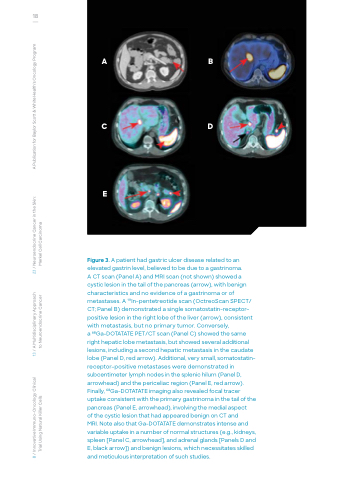
AB
CD
E
Figure 3. A patient had gastric ulcer disease related to an elevated gastrin level, believed to be due to a gastrinoma.
A CT scan (Panel A) and MRI scan (not shown) showed a cystic lesion in the tail of the pancreas (arrow), with benign characteristics and no evidence of a gastrinoma or of metastases. A 111In-pentetreotide scan (OctreoScan SPECT/ CT; Panel B) demonstrated a single somatostatin-receptor– positive lesion in the right lobe of the liver (arrow), consistent with metastasis, but no primary tumor. Conversely,
a 68Ga-DOTATATE PET/CT scan (Panel C) showed the same right hepatic lobe metastasis, but showed several additional lesions, including a second hepatic metastasis in the caudate lobe (Panel D, red arrow). Additional, very small, somatostatin- receptor–positive metastases were demonstrated in subcentimeter lymph nodes in the splenic hilum (Panel D, arrowhead) and the periceliac region (Panel E, red arrow). Finally, 68Ga-DOTATATE imaging also revealed focal tracer uptake consistent with the primary gastrinoma in the tail of the pancreas (Panel E, arrowhead), involving the medial aspect
of the cystic lesion that had appeared benign on CT and
MRI. Note also that Ga-DOTATATE demonstrates intense and variable uptake in a number of normal structures (e.g., kidneys, spleen [Panel C, arrowhead], and adrenal glands [Panels D and E, black arrow]) and benign lesions, which necessitates skilled and meticulous interpretation of such studies.
8 / Innovative Immuno-Oncology Clinical 13 / A Multidisciplinary Approach 23 / Neuroendocrine Cancer in the Skin: A Publication for Baylor Scott & White Health’s Oncology Program Trial Using Natural Killer Cells to Neuroendocrine Cancer Merkel Cell Carcinoma
