
Page 11 - Alaska A & P Primer
P. 11
Directional terms such as anterior and posterior are essential for accurately describing the relative locations of body structures. Images of the body’s interior commonly align along one of three planes: the sagittal, frontal, or transverse.
The body’s organs are organized in one of two main caviti- es—dorsal (also re- ferred to posterior) and ventral (also re- ferred to anteri- or)—which are fur- ther sub-divided ac- cording to the struc- tures present in each area.
The serous mem- branes have two lay- ers—parietal and vis- ceral—surrounding a fluid filled space. Serous membranes cover the lungs (pleu- ral serosa), heart (pericardial serosa), and some abdomin- opelvic organs (peri- toneal serosa).
1.7 Medical Imaging
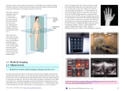
Detailed anatomical drawings of the human body first became available in the fifteenth and sixteenth centuries; however, it was not until the end of the nineteenth century, and the discovery of X-rays, that anatomists and physicians discovered non-surgical methods to look inside a living body. Since then, many other techniques, including CT scans, MRI scans, PET scans, and ultrasonography, have been developed, providing more accurate and detailed views of the form and function of the human body. In 1895, Rentgen made the first durable record of the internal parts of a living human: an “X-ray” image (as it
came to be called) of his wife’s hand. Scientists around
the world quickly began their own experiments with X-
rays, and by 1900, X-rays were widely used to detect a variety of injuries and diseases. (a) The results of a CT
scan of the head are shown as successive transverse sec- tions. (b) An MRI machine generates a magnetic field around a patient. (c) PET scans use radiopharmaceuticals
to create images of active blood flow and physiologic activ- ity of the organ or organs being targeted. (d) Ultrasound technology is used to monitor pregnancies because it is
the least invasive of imaging techniques and uses no elec- tromagnetic radiation. (credit a: Akira Ohgaki/flickr;
credit b: “Digital Cate”/flickr; credit c: “Raziel”/
Wikimedia Commons; credit d: “Isis”/Wikimedia Commons)
1.7 OBJECTIVES
1. Identify four modern medical imaging techniques and their uses
Watch this video (http://openstaxcollege.org/l/CATscan) to learn more about CT and CAT scans. What type of radiation does a CT
This content is available for free at https://cnx.org/content/col11496/1.7
State of Alaska EMS Education Primer - 2016
10
