
Page 117 - Alaska A & P Primer
P. 117
20.1 Structure and Function of Blood Vessels
Blood pumped by the heart flows through a series of vessels known as arteries, arterioles, capillaries, venules, and veins before returning to the heart. Arteries transport blood away from the heart and branch into smaller vessels, forming arterioles. Arterioles distribute blood to capillary beds, the sites of exchange with the body tissues. Capillaries lead back to small vessels known as venules that flow into the larger veins and eventually back to the heart.
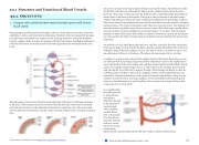
The pulmonary circuit moves blood from the right side of the heart to the lungs and back
to the heart. The systemic circuit moves blood from the left side of the heart to the head and body and returns it to the right side of the heart to repeat the cycle. The arrows indi- cate the direction of blood flow, and the colors show the relative levels of oxygen concentra- tion.
An artery is a blood vessel that conducts blood away from the heart. All arteries have rela- tively thick walls that can withstand the high pressure of blood ejected from the heart. However, those close to the heart have the thickest walls, containing a high percentage of elastic fibers in all three of their tunics. This type of artery is known as an elastic artery. Farther from the heart, where the surge of blood has dampened, the percentage of elastic fibers in an artery’s tunica intima decreases and the amount of smooth muscle in its tunica media increases. The artery at this point is described as a muscular artery. The diameter of muscular arteries typically ranges from 0.1 mm to 10 mm. Their thick tunica media allows muscular arteries to play a leading role in vasoconstriction. In contrast, their decreased quantity of elastic fibers limits their ability to expand. Fortunately, because the blood pres- sure has eased by the time it reaches these more distant vessels, elasticity has become less important.
An arteriole is a very small artery that leads to a capillary. Arterioles have the same three tunics as the larger vessels, but the thickness of each is greatly diminished. The critical en- dothelial lining of the tunica intima is intact. The tunica media is restricted to one or two smooth muscle cell layers in thickness. The tunica externa remains but is very thin.
A capillary is a microscopic channel that supplies blood to the tissues themselves, a proc- ess called perfusion. Exchange of gases and other substances occurs in the capillaries be- tween the blood and the surrounding cells and their tissue fluid (interstitial fluid). The di- ameter of a capillary lumen ranges from 5–10 micrometers; the smallest are just barely wide enough for an erythrocyte to squeeze through. Flow through capillaries is often de- scribed as microcirculation. The wall of a capillary consists of the endothelial layer sur- rounded by a basement membrane with occasional smooth muscle fibers. There is some variation in wall structure: In a large capillary, several endothelial cells bordering each other may line the lumen; in a small capillary, there may be only a single cell layer that wraps around to contact itself.
In a capillary bed,
arterioles give rise
to metarterioles.
Precapillary
sphincters located
at the junction of
a metarteriole
with a capillary
regulate blood
flow. A thorough-
fare channel con-
nects the metarte-
riole to a venule.
An arteriovenous
anastomosis,
which directly connects the arteriole with the venule, is shown at the bottom.
20.1 OBJECTIVES
1. Compare and contrast the three tunics that make up the walls of most blood vessels
This content is available for free at https://cnx.org/content/col11496/1.7
State of Alaska EMS Education Primer - 2016
116
