
Page 121 - Alaska A & P Primer
P. 121
to clot the blood. This clot can further obstruct the artery and—if it occurs in a coronary or cerebral artery—cause a sudden heart attack or stroke. Alternatively, plaque can break off and travel through the bloodstream as an embolus until it blocks a more distant, smaller artery.
Even without total blockage, vessel narrowing leads to ischemia—reduced blood flow—to the tissue region “downstream” of the narrowed vessel. Ischemia in turn leads to hy- poxia—decreased supply of oxygen to the tissues. Hypoxia involving cardiac muscle or brain tissue can lead to cell death and severe impairment of brain or heart function.
A major risk factor for both arteriosclerosis and atherosclerosis is advanced age, as the con- ditions tend to progress over time. Arteriosclerosis is normally defined as the more general- ized loss of compliance, “hardening of the arteries,” whereas atherosclerosis is a more spe- cific term for the build-up of plaque in the walls of the vessel and is a specific type of arte- riosclerosis. There is also a distinct genetic component, and pre-existing hypertension and/or diabetes also greatly increase the risk. However, obesity, poor nutrition, lack of physical activity, and tobacco use all are major risk factors.
Treatment includes lifestyle changes, such as weight loss, smoking cessation, regular exer- cise, and adoption of a diet low in sodium and saturated fats. Medications to reduce choles- terol and blood pressure may be prescribed. For blocked coronary arteries, surgery is war- ranted. In angioplasty, a catheter is inserted into the vessel at the point of narrowing, and
a second catheter with a balloon-like tip is inflated to widen the opening. To prevent subse- quent collapse of the vessel, a small mesh tube called a stent is often inserted. In an en- darterectomy, plaque is surgically removed from the walls of a vessel. This operation is typically performed on the carotid arteries of the neck, which are a prime source of oxygen- ated blood for the brain. In a coronary bypass procedure, a non-vital superficial vessel from another part of the body (often the great saphenous vein) or a synthetic vessel is in- serted to create a path around the blocked area of a coronary artery.
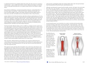
The contraction of skeletal muscles surrounding a vein compresses the blood and in- creases the pressure in that area. This action forces blood closer to the heart where venous pressure is lower. Note the importance of the one-way valves to assure that blood flows only in the proper direction.
The respiratory pump aids blood flow through the veins of the thorax and abdomen. Dur- ing inhalation, the volume of the thorax increases, largely through the contraction of the diaphragm, which moves downward and compresses the abdominal cavity. The elevation of the chest caused by the contraction of the external intercostal muscles also contributes to the increased volume of the thorax. The volume increase causes air pressure within the thorax to decrease, allowing us to inhale. Additionally, as air pressure within the thorax drops, blood pressure in the thoracic veins also decreases, falling below the pressure in the abdominal veins. This causes blood to flow along its pressure gradient from veins outside the thorax, where pressure is higher, into the thoracic region, where pressure is now lower. This in turn promotes the return of blood from the thoracic veins to the atria. During exha- lation, when air pressure increases within the thoracic cavity, pressure in the thoracic
veins increases, speeding blood flow into the heart while valves in the veins prevent blood from flowing backward from the thoracic and abdominal veins.
Although vessel diameter increases from the smaller venules to the larger veins and eventu- ally to the venae cavae (singular = vena cava), the total cross-sectional area actually de- creases. The individual veins are larger in diameter than the venules, but their total num- ber is much lower, so their total cross-sectional area is also lower. Also notice that, as blood moves from venules to veins, the average blood pressure drops, but the blood veloc- ity actually increases. This pressure gradient drives blood back toward the heart. Again, the presence of one-way valves and the skeletal muscle and respiratory pumps contribute to this increased flow. Since approximately 64 percent of the total blood volume resides in systemic veins, any action that increases the flow of blood through the veins will increase venous return to the heart. Maintaining vascular tone within the veins prevents the veins from merely distending, dampening the flow of blood, and as you will see, vasoconstriction actually enhances the flow.
As previously discussed, vasoconstriction of an artery or arteriole decreases the radius, increasing resistance and pressure, but decreasing flow. Venoconstriction, on the other hand, has a very different outcome. The walls of veins are thin but irregular; thus, when the smooth muscle in those walls constricts, the lumen becomes more rounded. The more rounded the lumen, the less surface area the blood encounters, and the less resistance the vessel offers. Vasoconstriction increases pressure within a vein as it does in an artery, but in veins, the increased pressure increases flow.
Recall that the pres- sure in the atria, into which the venous blood will flow, is very low, approach- ing zero for at least part of the relaxation phase of the cardiac cycle. Thus, venocon- striction increases the return of blood to the heart.
Another way of stat- ing this is that veno- constriction in- creases the preload or stretch of the car- diac muscle and in- creases contraction.
This content is available for free at https://cnx.org/content/col11496/1.7
State of Alaska EMS Education Primer - 2016
120
