
Page 124 - Alaska A & P Primer
P. 124
the arteries that supplied these regions and the two often parallel one another. This is of- ten described as a “complementary” pattern. However, there is a great deal more variabil- ity in the venous circulation than normally occurs in the arteries. For the sake of brevity and clarity, this text will discuss only the most commonly encountered patterns. However, keep this variation in mind when you move from the classroom to clinical practice.
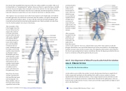
The superior vena cava drains most of the body superior to the diaphragm. On both the left and right sides, the subclavian vein forms when the axillary vein passes through the body wall from the axillary region. It fuses with the external and internal jugular veins from the head and neck to form the brachiocephalic vein. Each vertebral vein also flows into the brachiocephalic vein close to this fusion.
The remainder of the blood supply from the thorax drains into the azygos vein. Each in- tercostal vein drains muscles ofthe thoracic wall, each esophageal vein delivers blood from the inferior por- tions of the esopha- gus, each bronchial veindrains the sys- temic circulation from the lungs, and several smaller veins drain the mediastinal region. Bronchial veins carry approximately 13 per- cent of the blood that flows into the bron- chial arteries; the re- mainder intermingles with the pulmonary circulation and re- turns to the heart via the pulmonary veins.
Blood from the brain and the superficial fa- cial vein flow into each internal jugular vein . Many smaller veins of the brain stem and the superficial veins of the
cerebrum lead to larger vessels referred to as intracranial si- nuses. These in- clude the supe- rior and inferior sagittal sinuses, straight sinus, cavernous si- nuses, left and right sinuses, the petrosal sinuses, and the occipital sinuses.
After oxygenat-
ing tissues in the
capillaries, sys-
temic blood is
returned to the
right atrium
from the venous
system via the superior vena cava, which drains most of the veins superior to the dia- phragm, the inferior vena cava,which drains most of the veins inferior to the diaphragm, and the coronary veins via the coronary sinus. The hepatic portal system carries blood to the liver for processing before it enters circulation. Review the figures provided in this sec- tion for circulation of blood through the blood vessels.
20.6 Development of Blood Vessels adn Fetal Circulation
As the embryo grows within the mother’s womb, the placenta develops to supply blood
rich in oxygen and nutrients via the umbilical vein and to remove wastes in oxygen- depleted blood via the umbilical arteries. Three major shunts found in the fetus are the foramen ovale and ductus arteriosus, which divert blood from the pulmonary to the sys- temic circuit, and the ductus venosus, which carries freshly oxygenated blood high in nutri- ents to the fetal heart.
20.6 OBJECTIVES
1. Describe the fetal circulation
This content is available for free at https://cnx.org/content/col11496/1.7
State of Alaska EMS Education Primer - 2016
123
