
Page 128 - Alaska A & P Primer
P. 128
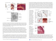
(a) The spleen is attached to the stomach. (b) A micrograph of spleen tissue shows the ger- minal center. The marginal zone is the region between the red pulp and white pulp, which sequesters particulate antigens from the circulation and presents these antigens to lympho- cytes in the white pulp. EM Å~ 660. (Micrograph provided by the Regents of the Univer- sity of Michigan Medical School ˝ 2012)
In addition to the lymph nodes, the spleen is a major secondary lymphoid organ. It is about 12 cm (5 in) long and is attached to the lateral border of the stomach via the gas- trosplenic ligament. The spleen is a fragile organ without a strong capsule, and is dark red due to its extensive vascularization. The spleen is sometimes called the “filter of the blood” because of its extensive vascularization and the presence of macrophages and dendritic cells that remove microbes and other materials from the blood, including dying red blood cells. The spleen also functions as the location of immune responses to blood-borne patho- gens.
Tonsils are lymphoid nodules located along the inner surface of the pharynx and are impor- tant in developing immunity to oral pathogens. The tonsil located at the back of the throat, the pharyngeal tonsil, is sometimes referred to as the adenoid when swollen. Such swelling is an indication of an active immune response to infection. Histologically, tonsils do not contain a complete capsule, and the epithelial layer invaginates deeply into the interior of
the tonsil to form tonsillar crypts. These structures, which accumulate all sorts of materi- als taken into the body through eating and breathing, actually “encourage” pathogens to penetrate deep into the tonsillar tissues where they are acted upon by numerous lymphoid follicles and eliminated. This seems to be the major function of tonsils—to help children’s bodies recognize, destroy, and develop immunity to common environmental pathogens so that they will be protected in their later lives. Tonsils are often removed in those children who have recurring throat infections, especially those involving the palatine tonsils on ei- ther side of the throat, whose swelling may interfere with their breathing and/or swallow- ing.
The pharyngeal tonsil is located on the roof of the posterior supe- rior wall of the nasopharynx. The palatine ton- sils lay on each side of the phar- ynx. (b) A micro- graph shows the palatine tonsil tissue. LM X 40. (Micrograph pro- vided by the Re- gents of the Uni- versity of Michi- gan Medical School ˝ 2012)
Mucosa-
associated lym-
phoid tissue
(MALT) consists
of an aggregate of
lymphoid folli-
cles directly asso-
ciated with the
mucous mem-
brane epithelia.
MALT makes up
dome-shaped structures found underlying the mucosa of the gastrointestinal tract, breast tissue, lungs, and eyes. Peyer’s patches, a type of MALT in the small intestine, are espe- cially important for immune responses against ingested substances. Bronchus-associated lymphoid tissue (BALT) consists of lymphoid follicular structures with an overlying epithe- lial layer found along the bifurcations of the bronchi, and between bronchi and arteries.
This content is available for free at https://cnx.org/content/col11496/1.7
State of Alaska EMS Education Primer - 2016
127
