
Page 53 - Alaska A & P Primer
P. 53
9.4 Synovial Joints
Bursae are classified by their location.
Bursitis
http://www.michiganpain.com/bursitis.php
9.4 OBJECTIVES
1. Describe the structural features of the six types of synovial joints
Synovial joints are characterized by the presence of a joint cavity. The walls of this space are formed by the articular capsule, a fibrous connective tissue structure that is attached to each bone just outside the area of the
bone’s articulating surface.
Bursitis is the inflammation of a bursa near a joint. This will cause pain, swelling, or ten- derness of the bursa and surrounding area, and may also result in joint stiffness.
Bursitis is most commonly associated with the bursae found at or near the shoulder, hip, knee, or elbow joints. At the shoulder, subacromial bursitis may occur in the bursa that separates the acromion of the scapula from the tendon of a shoulder muscle as it passes deep to the acromion. In the hip region, trochanteric bursitis can occur in the bursa that overlies the greater trochanter of the femur, just below the lateral side of the hip. Ischial bursitis occurs in the bursa that separates the skin from the ischial tuberosity of the pel- vis, the bony structure that is weight bearing when sitting. At the knee, inflammation and swelling of the bursa located between the skin and patella bone is prepatellar bursitis (“housemaid’s knee”), a condition more commonly seen today in roofers or floor and car- pet installers who do not use knee pads. At the elbow, olecranon bursitis is inflammation of the bursa between the skin and olecranon process of the ulna.
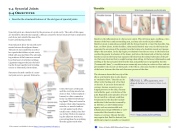
Synovial joints allow for smooth move- ments between the adjacent bones. The joint is surrounded by an articu- lar capsule that defines a joint cavity filled with synovial fluid. The articulat- ing surfaces of the bones are covered by a thin layer of articular cartilage. Ligaments support the joint by hold- ing the bones together and resisting excess or abnormal joint motions.
Structures located outside of a syno- vial joint serve to prevent friction be-
The olecranon forms the bony tip of the elbow, and bursitis here is also known as “student’s elbow.” Bursitis can be either acute (lasting only a few days)
or chronic. It can arise from muscle overuse, trauma, excessive or pro-
longed pressure on the skin, rheuma- toid arthritis, gout, or infection of the joint. Repeated acute episodes of bursi- tis can result in a chronic condition. Treatments for the disorder include antibiotics if the bursitis is caused by
an infection, or anti-inflammatory agents, such as nonsteroidal anti- inflammatory drugs (NSAIDs) or corti- costeroids if the bursitis is due to
trauma or overuse. Chronic bursitis
may require that fluid be drained, but additional surgery is usually not required.
tween the bones of the joint and the overlying muscle ten- dons or skin. A bursa (plural = bursae) is a thin connective tissue sac filled with lubricat- ing liquid. They are located in regions where skin, ligaments, muscles, or muscle tendons can rub against each other, usually near a body joint. Bur- sae reduce friction by separat- ing the adjacent structures, preventing them from rubbing directly against each other.
MOVIE 1.18 Ligaments, Ten- dons & Joints 2:47 minutes, Khan Acad- emy
Watch https://youtu.be/frvv37WJjm4
This content is available for free at https://cnx.org/content/col11496/1.7
State of Alaska EMS Education Primer - 2016
52
