
Page 73 - Alaska A & P Primer
P. 73
The thigh muscles that move the femur, tibia, and fibula are divided into medial, anterior, and posterior compartments. The medial compartment includes the adductors, pectineus, and the gracilis. The anterior compartment comprises the quadriceps femoris, quadriceps tendon, patellar ligament, and the sartorius. The quadriceps femoris is made of four mus- cles: the rectus femoris, the vastus lateralis, the vastus medius, and the vastus interme- dius, which together extend the knee. The posterior compartment of the thigh includes the hamstrings: the biceps femoris, semitendinosus, and the semimembranosus, which all flex the knee. The muscles of the leg that move the foot and toes are divided into anterior, lat- eral, superficial- and deep-posterior compartments.
The muscles of the anterior compartment of the lower leg are generally responsible for dor- siflexion, and the muscles of the posterior compartment of the lower leg are generally re- sponsible for plantar flexion. The lateral and medial muscles in both compartments invert, evert, and rotate the foot.
The posterior compartment of the thigh includes muscles that flex the leg and extend the thigh. The three long muscles on the back of the knee are the hamstring group, which flexes the knee. These are the biceps femoris, semitendinosus, and semimembranosus. The tendons of these muscles form the popliteal fossa, the diamond-shaped space at the back of the knee.
The posterior compartment of the thigh includes muscles that flex the leg and extend the thigh. The three long muscles on the back of the knee are the hamstring group, which flexes the knee. These are the biceps femoris, semitendinosus, and semimembranosus. The tendons of these muscles form the popliteal fossa, the diamond-shaped space at the back of the knee.
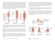
The muscles along the dorsal side of the foot (a) generally extend the toes while the mus- cles of the plantar side of the foot (b, c, d) generally flex the toes. The plantar muscles exist in three layers, providing the foot the strength to counterbalance the weight of the body. In this diagram, these three layers are shown from a plantar view beginning with the bottom- most layer just under the plantar skin of the foot (b) and ending with the top-most layer (d) located just inferior to the foot and toe bones.
The foot also has intrinsic muscles, which originate and insert within it (similar to the in- trinsic muscles of the hand). These muscles primarily provide support for the foot and its arch, and contribute to movements of the toes.
The principal support for the longitudinal arch of the foot is a deep fascia called plantar aponeurosis, which runs from the calcaneus bone to the toes (inflammation of this tissue is the cause of “plantar fasciitis,” which can affect runners. The intrinsic muscles of the foot consist of two groups. The dorsal group includes only one muscle, the extensor digitorum brevis. The second group is the plantar group, which consists of four layers, starting with the most superficial.
This content is available for free at https://cnx.org/content/col11496/1.7
State of Alaska EMS Education Primer - 2016
72
