
Page 76 - Alaska A & P Primer
P. 76
12.2 Nervous Tissue
12.3 The Function of Nervous Tissue
12.2 OBJECTIVES
1. Describe the basic structure of a neuron
12.3 OBJECTIVES
1. Distinguish the major functions of the nervous system: sensation, integration, and response
The picture you have in your mind of the nervous system probably includes the brain, the nervous tissue contained within the cranium, and the spinal cord, the extension of nervous tissue within the vertebral column. That suggests it is made of two organs—and you may not even think of the spinal cord as an organ—but the nervous system is a very complex structure. Within the brain, many different and separate regions are responsible for many different and separate functions. It is as if the nervous system is composed of many organs that all look similar and can only be differentiated using tools such as the microscope or electrophysiology.
In comparison, it is easy to see that the stomach is different than the esophagus or the liver, so you can imagine the digestive system as a collection of specific organs. Neurons are the cells considered to be the basis of nervous tissue. They are responsible for the elec- trical signals that communicate information about sensations, and that produce move- ments in response to those stimuli, along with inducing thought processes within the brain.
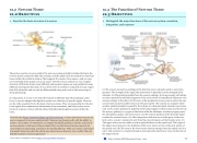
(1) The sensory neuron has endings in the skin that sense a stimulus such as water tem- perature. The strength of the signal that starts here is dependent on the strength of the stimulus. (2) The graded potential from the sensory endings, if strong enough, will initiate an action potential at the initial segment of the axon (which is immediately adjacent to the sensory endings in the skin). (3) The axon of the peripheral sensory neuron enters the spi- nal cord and contacts another neuron in the gray matter. The contact is a synapse where another graded potential is caused by the release of a chemical signal from the axon termi- nals. (4) An action potential is initiated at the initial segment of this neuron and travels up the sensory pathway to a region of the brain called the thalamus. Another synapse passes the information along to the next neuron. (5) The sensory pathway ends when the signal reaches the cerebral cortex. (6) After integration with neurons in other parts of the cere- bral cortex, a motor command is sent from the precentral gyrus of the frontal cortex. (7) The upper motor neuron sends an action potential down to the spinal cord. The target of the upper motor neuron is the dendrites of the lower motor neuron in the gray matter of the spinal cord. (8) The axon of the lower motor neuron emerges from the spinal cord in a nerve and connects to a muscle through a neuromuscular junction to cause contraction of the target muscle.
Visit this site (http://openstaxcollege.org/l/nervetissue3) to learn about how nervous tis- sue is composed of neurons and glial cells. Neurons are dynamic cells with the ability to make a vast number of connections, to respond incredibly quickly to stimuli, and to initi- ate movements on the basis of those stimuli. They are the focus of intense research be- cause failures in physiology can lead to devastating illnesses. Why are neurons only found in animals? Based on what this article says about neuron function, why wouldn't they be helpful for plants or microorganisms?
This content is available for free at https://cnx.org/content/col11496/1.7
State of Alaska EMS Education Primer - 2016
75
