
Page 81 - Alaska A & P Primer
P. 81
13.3 Circulation and the Central Nervous System
13.3 OBJECTIVES
1. Describe the vessels that supply the CNS with blood
The CNS has a privileged blood supply, as suggested by the blood-brain barrier. The func- tion of the tissue in the CNS is crucial to the survival of the organism, so the contents of the blood cannot simply pass into the central nervous tissue.
To protect this region from the toxins and pathogens that may be traveling through the blood stream, there is strictcontrol over what can move out of the general systems and into the brain and spinal cord. Because of this privilege, the CNS needs specialized structures for the maintenance of circulation. This begins with a unique arrangement of blood vessels carrying fresh blood into the CNS. Beyond the supply of blood, the CNS filters that blood into cerebrospinal fluid (CSF),
which is then circulated through the
cavities of the brain and spinal cord
called ventricles.
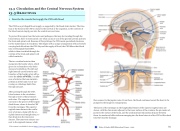
The two vertebral arteries then merge into the basilar artery, which gives rise to branches to the brain stem and cerebellum. The left and right internal carotid arteries and branches of the basilar artery all be- come the circle of Willis, a conflu- ence of arteries that can maintain perfusion of the brain even if nar- rowing or a blockage limits flow through one part.
After passing through the CNS, blood returns to the circulation through a series of dural sinuses
and veins. The superior sagittal si- nus runs in the groove of the longitu- dinal fissure, where it absorbs CSF from the meninges. The superior sagittal sinus drains to the conflu- ence of sinuses, along with the oc- cipital sinuses and straight sinus, to then drain into the transverse sinuses. The transverse sinuses con- nect to the sigmoid sinuses, which
then connect to the jugular veins. From there, the blood continues toward the heart to be pumped to the lungs for reoxygenation.
The layers of the meninges in the longitudinal fissure of the superior sagittal sinus are shown, with the dura mater adjacent to the inner surface of the cranium, the pia mater ad- jacent to the surface of the brain, and the arachnoid and subarachnoid space between them. An arachnoid villus isshown emerging into the dural sinus to allow CSF to filter back into the blood for drainage.
This content is available for free at https://cnx.org/content/col11496/1.7
State of Alaska EMS Education Primer - 2016
80
