
Page 78 - ro membanes
P. 78
3.4 MEMBRANE AUTOPSY 61
However, the tested tail element A608298 has been exposed to both excessive flux loss (37%) and extremely high irreversible salt passage increase of 144%, and therefore is not suit- able for rotation to lead position. As a consequence, this membrane element has to be replaced. As shown on Table 3.3, the results from the vacuum test indicate that this element cannot hold vacuum (e.g., it is irreversibly damaged), which confirms the observation from the cell test that the element is beyond repair and would need to be replaced.
3.4.9 SEM
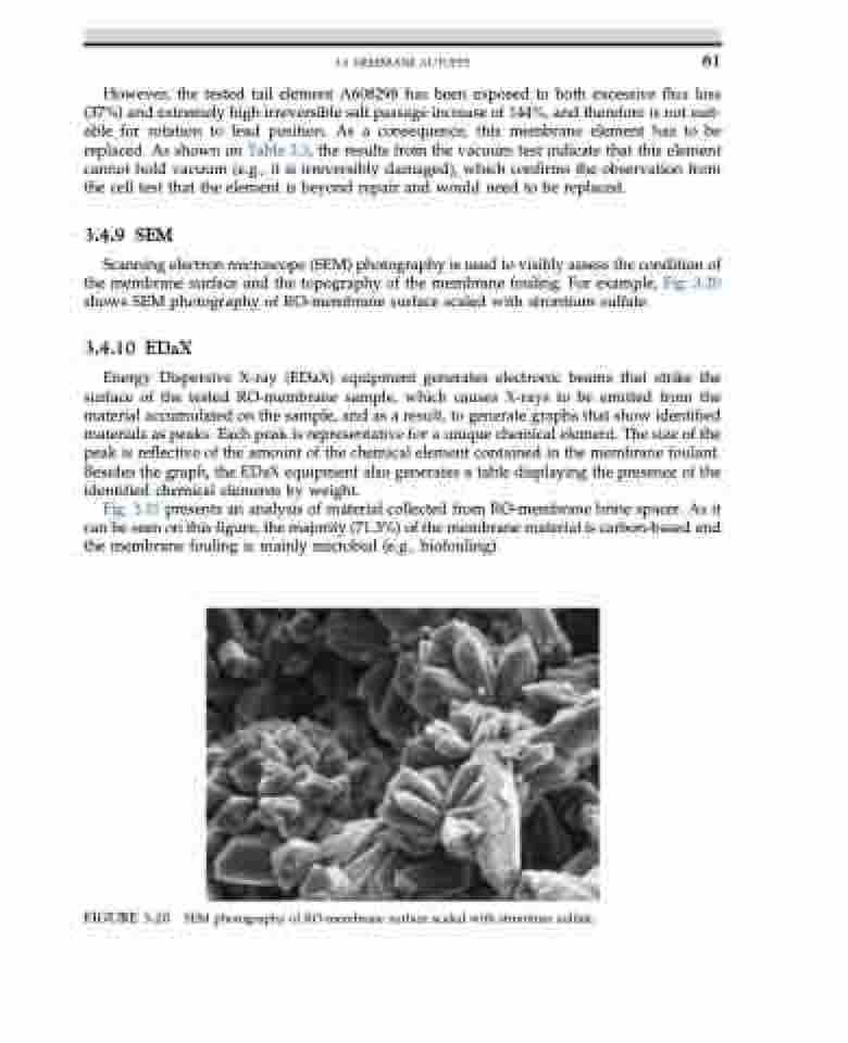
Scanning electron microscope (SEM) photography is used to visibly assess the condition of the membrane surface and the topography of the membrane fouling. For example, Fig. 3.20 shows SEM photography of RO-membrane surface scaled with strontium sulfate.
3.4.10 EDaX
Energy Dispersive X-ray (EDaX) equipment generates electronic beams that strike the surface of the tested RO-membrane sample, which causes X-rays to be emitted from the material accumulated on the sample, and as a result, to generate graphs that show identified materials as peaks. Each peak is representative for a unique chemical element. The size of the peak is reflective of the amount of the chemical element contained in the membrane foulant. Besides the graph, the EDaX equipment also generates a table displaying the presence of the identified chemical elements by weight.
Fig. 3.21 presents an analysis of material collected from RO-membrane brine spacer. As it can be seen on this figure, the majority (71.3%) of the membrane material is carbon-based and the membrane fouling is mainly microbial (e.g., biofouling).
FIGURE 3.20 SEM photography of RO-membrane surface scaled with strontium sulfate.
