
Page 104 - March_2022
P. 104
EQUINE HEALTH
STEM CELL
T T H H E E R R A A P P Y Y F F O OR R OSTEOARTHRITIS
Story by Heather Smith Thomas Photos provided by Karen Mantel, Ontario Veterinary College, University of Guelph and Dr. Erin Roberts, University of Calgary
Stem cell therapy has been utilized in horses to help heal tendon, ligament and joint injuries for more than 20 years, and new uses are continually explored. Stem cells used in horses are mesenchymal stromal cells (MSCs)
which are isolated from fetuses, foals or adult horses, as opposed to embryonic stem cells isolated from embryos. The MSC equine stem cells can be isolated from almost any tissue but most commonly obtained from bone
marrow, fat tissue and from the umbilical cord of newborn foals.
The use of certain stem cells requires
a laboratory culture to isolate them and expand their numbers. These cells are char- acterized by their ability to become other types of cells in the lab and to modify the function of cells from the immune system such as lymphocytes. Cells from bone mar- row and fat tissue are also used after a brief laboratory processing step where all the cells are concentrated from the sample without a culture to select and expand MSCs. These processes are known as bone marrow concen- trate or stromal vascular fraction, depending on whether the starting material is bone mar- row aspirate or fat tissue. Sometimes these cell preparations are referred to as stem cell treatments although very few of these cells would be MSCs.
Autologous versus allogeneic use of cells is another important distinction. Autologous cells are from the patient itself. Allogeneic cells are from a donor horse and are placed in other horses. The advantage of using autologous cells is that they are not rejected by the patient’s immune system, and there are less regulatory concerns with their use.
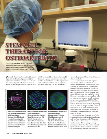
Equine Cord Blood Mesenchymal Stem Cells grown on Microcarriers in 0.1L Vertical Wheel Bioreactors from PBS Biotech. Several cells are attached to
the microcarrier bead. Cells are stained to show the cell nucleus in turquoise and actin filaments within the cells in pink.
Equine Cord Blood Mesenchymal Stem Cells grown on Microcarriers in 0.1L Vertical Wheel Bioreactors from PBS Biotech. Several cells are attached to the microcarrier bead. Cells are stained to show the cell nucleus in green and the cell membrane in blue.
Equine Cord Blood Mesenchymal Stem Cells grown on Microcarriers in 0/1L Vertical Wheel Bioreactors from PB Biotech. Several cells are attached to the microcarrier bead. Cells are stained to show the cell nucleus in blue and actin filaments within the cells in green.
102 SPEEDHORSE March 2022
Photos by Dr. Erin Roberts, University of Calgary
