
Page 100 - Speedhorse June 2020
P. 100
EQUINE HEALTH
may be difficult to repair and can end up with scar tissue that comes over the side of the col- lateral cartilage and coronary band.
“I’ve also seen foreign bodies that penetrate into the coronary band,” says Peters. “I’ve seen horses with splinters from a fence embedded deeply into those tissues, or a piece of wood/ tree branch jammed down inside the hoof wall, right through the coronary band. This is like driving a nail under your toenail or fingernail. These can be very sore, and those splinters can sometimes puncture into the coffin joint itself and be serious.”
TREATMENT AND REPAIR
Dr. Olivia Rudolphi of Rudolphi Veterinary Services, in Noble, Illinois, says the coronary band injuries she sees are often lacerations from getting a foot caught. “This may involve a cut clear through the coronary band or a flap that pulls downward and pulls the hoof wall off with it, or flips upward and still has the skin attached,” she explains.
“Depending on the site of the injury, a lac- eration can sometimes be closed with sutures, if adequate tissue still remains,” Rudophi con- tinues. “Sometimes we have to just remove that entire portion that is unattached. We also need to make sure there’s not a puncture going into the coffin joint or pastern joint. It’s wise to use antibiotics and make sure the horse’s tetanus vaccinations are up to date.
“We have to make sure the coffin joint is not infected, and then if we can suture it, we will. If we can’t, we may let it heal by second intention (with the wound healing from the inside out). We may use some biologic or regenerative thera- pies to help it grow back a little bit better.”Some of these injuries will need careful evaluation and surgical repair. “There are many important structures right around the coronary band, in- cluding the coffin joint,” says Peters. “The bony recesses/pouches of this joint come up pretty close to the collateral cartilages that come off the coffin bone. These can be damaged if some- thing cuts into them, and can potentially cause infections. The bones themselves—the pastern and coffin bone—can be injured, along with their ligament attachments. This may include the coffin joint and pastern joints.”
Careful evaluation of any damage or lacera- tion is imperative to see what structures may be involved. “If there is joint fluid present in the in- jury, we will probably need to flush those joints and keep that lower leg area bandaged and very clean. The treatment may be quite complicated in order to have a good outcome,” Peters says.
Cassells says that once the initial care is accomplished (immediately after the injury, applying pressure and stop the bleeding), it’s important to clean the hoof and get it properly
bandaged and dressed to allow that area to heal. “This is a difficult area to stitch, partly because there is a lot of movement but also be- cause that tissue does not hold suture material very well. If it is right on the coronary band, we can’t suture into the hoof wall. Usually we depend on appropriate bandaging to stabilize the area, and let it heal by what we call second intention (as opposed to sutures). This means we simply let the wound close over from the inside out. This is usually the best way to get these to heal, just because they are often dif- ficult to stitch. Obviously, if the wound comes farther up onto the pastern, we can suture that area,” he explains.
Sometimes a cast is used to immobilize
the tissues until they can heal and grow back together. “If we’re going to apply a cast, the area has to be adequately cleaned first because, otherwise, it will just trap infection in there. Some people prefer to use a cast since this simplifies the care, and it’s not as involved as
a daily bandage change. There are different schools of thought on this. Some people like to take the bandage off every day and take
a look at it to make sure it’s healing the way they want; other people want to put a cast on, as a low-maintenance treatment option. It all depends on the situation, the horse, the owner, and the veterinarian who is in charge of the case,” Cassells says.
“I have found that sometimes it’s a good idea to take a couple of radiographs just to make sure there is no deeper damage, such as to the coffin bone or joint. In most cases these wounds don’t get that deep, so x-rays may or may not be warranted, based on the case. The times I usually try to take an x-ray are when the horse is very lame—much more lame than it should be for just a wound, or if there is injury from a foreign object such as sheet metal if the horse kicked through the barn wall,” he says.
Sometimes a horse may jam a foreign
object in through the coronary band, such as a splinter of wood. If there is suspicion that part of that foreign object may still be embedded into the hoof, it’s good to do some imaging like an x-ray or ultrasound just to make sure there are no fragments still inside. In those cases, the fragments would need to be surgically removed, and this can be a challenge.
“It can be very rewarding, however, when we get it out of there, like getting the splinter out from under your fingernail. Antibiotics are recommended for these situations, and usually any time there is a deep wound to the coronary band, because the foot is always in an unsani- tary environment, whether walking around in the stall, pen or pasture. There’s always some dust and dirt, so antibiotics are a good idea,” says Cassells.
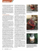
Coronary band injuries are often traumatic and include severe lacerations.
A laceration can sometimes be closed with sutures, depending on the site of the injury and if adequate tissue remains.
It can take anywhere from 10 to 12 months for some coronary band injuries to completely grow a new hoof capsule, possibly leaving a hoof
wall defect or weakness that may be a potential problem for later performance.
98 SPEEDHORSE June 2020
Hailie DeVries Hailie DeVries
