
Page 84 - December 2021
P. 84
EQUINE HEALTH
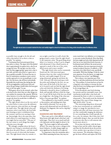
The right dorsal colon is tucked in behind the liver and readily imaged to check the thickness of its lining, which should be about 2 millimeters thick.
as possible (just enough to do the job and relieve discomfort) for as short a time as possible,” he explains.
“Veterinarians have been pretty good about
their advice to clients. The upper end of the labeled dose is approximately two grams twice a day for an average size horse. We may give one dose at this level, but then suggest giving 1 gram twice a day to start with, and then wean down to 1 gram once a day as quickly as possible. For horses that stay on bute for maintenance soundness or pain control, another approach is to not necessarily give it every day. Give the horse a day off from it periodically. Essentially this would mean that during that day off, the prostaglandins could bounce back and restore the gut lining to health before the horse goes back on the bute again,” he says.
“With gastric ulcers (in the stomach, rather than so far back in the colon) there are other things you can combine for treatment, like antacids. With the colon, however, it is so far away from anything you can give the horse by mouth that it is difficult to protect it,” he explains.
“The right dorsal colon is at the very end of the colon before it turns across the abdomen as the transverse colon and then out through the small colon. The right dorsal colon and transverse colon is just before the fecal balls start forming as water is withdrawn from
the contents, and feces are starting to firm
up. Interestingly, this is the same place that sand collects in a sand impaction, or where you might find an enterolith.” This part of the tract seems to catch things that might otherwise move through.
“We jokingly complain about the horse’s digestive tract being so convoluted and often talk about the pelvic flexure—and why would there be an area of colon with this hairpin turn. The right dorsal colon, by contrast, is
not as tight a turn but it is still a bend, like going around a corner, from the right dorsal to the transverse colon. The good thing about where it is, however, is that it can be imaged with ultrasound. It has a fixed location, as opposed to much of the rest of the colon, which moves and is more variable.”
“The right dorsal colon can be consistently found near the 12th intercostal space (between those two ribs), tucked in behind the liver, and readily imaged. If we are
using ultrasound--going up and down the 11th, 12th and 13th intercostal space with
the probe and looking for the liver, we can then come off the back of the liver once
we’ve located it and can find the right dorsal colon and check the thickness of its lining. Ordinarily it should be about 2 millimeters thick, and typically will have a little bit of gas in it. So just off the back of the liver you would see a bright line—the lining of the colon just before it becomes gas-filled,” he explains.
“Two millimeters or less would be normal,
at that position, and anything more than
that would be abnormal. You can see the mucosa with ultrasound and can tell when it’s thickened. For diagnosis, this can be a big help,” says Blikslager.
DIAGNOSIS AND TREATMENT
“Most cases can be a little difficult to pick up. Most horses with right dorsal colitis have only subtle signs. They won’t eat as much and look
a bit depressed, and this might be about it, for signs.” The horse might be dull and possibly
off feed. The owner may notice a change in the horse’s behavior. A subtle change might not seem significant but should not be ignored.
“It is important to have the veterinarian take a look. When examining the horse, they may not
notice much that’s very definitive, so it is important to run some routine bloodwork. More than likely the horse might just look a little depressed and off feed, but we can check blood levels of protein. A biochemical panel will show the serum protein
and the albumen, the most important protein for normal circulatory function. In addition, a CBC may show a low white blood count, specifically neutrophils. With these findings, I’d ask the owner more questions. From the history, we might learn that the horse was on bute,” says Blikslager.
This is always a very important part of the history, to ask what medications the horse has been on. “One problem, if a horse has been on bute for a long time, is the tendency to increase the dose when the horse is not feeling as good, which would be the wrong thing to do.
“If the bloodwork came back showing
low protein and low neutrophil count, your veterinarian would either come back and ultrasound the horse or refer it to someone to ultrasound it. This would reveal the thickened gut lining, if the cause of these changes was right dorsal colitis,” he says.
“The interesting thing about ultrasound
is that most veterinarians are comfortable using it to image the reproductive tract, tendons and other soft tissue structures, but not that comfortable imaging the abdomen. It’s not something they would typically do for a colic, for instance. But if they don’t feel comfortable doing it, they can send the horse to someone who does or call someone who knows more about it.
“If you have the history, with the horse being on bute and the subtle clinical signs, and then the bloodwork shows the protein is low, another way to go about it is just to start treatment. The immediate thing is to get the horse off bute and replace the PGE
82 SPEEDHORSE December 2021
