
Page 116 - Libro 2
P. 116
96
PART 2 — CEREBROVASCULAR
ABC
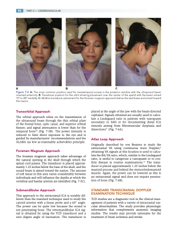
Figure 7-5 A: The most common position used for transtemporal access is the posterior window with the ultrasound beam oriented anteriorly. B: Transducer position for the orbit showing placement over the center of the eyelid with the beam aimed 15° to 20° medially. C: Midline transducer placement for the foramen magnum approach below the skull base and aimed toward the nasion.
Transorbital Approach
The orbital approach relies on the transmission of the ultrasound beam through the thin orbital plate of the frontal bone, optic canal, and superior orbital fissure; and signal attenuation is lower than for the temporal bone10 (Fig. 7-5B). The power intensity is reduced to limit direct exposure to the eye and is guided by manufacturers’ recommendations and the ALARA (as low as reasonably achievable) principle.
Foramen Magnum Approach
The foramen magnum approach takes advantage of the natural opening in the skull through which the spinal cord passes. The transducer is placed approxi- mately 1.25 inches below the base of the skull, and the sound beam is aimed toward the nasion. The amount of soft tissue in this area varies considerably between individuals and will influence the depths at which the vertebral and basilar arteries are identified (Fig. 7-5C).
Submandibular Approach
This approach to the extracranial ICA is notably dif- ferent than the standard technique used to study the carotid arteries with a linear probe and a 60° angle. The power can be quite low because the sound is not penetrating bone. The retromandibular ICA sig- nal is obtained by using the TCD transducer and a zero degree angle of insonation. The transducer is
placed at the angle of the jaw with the beam-directed cephalad. Signals obtained are usually used to calcu- late a Lindegaard ratio in patients with vasospasm secondary to SAH or for documenting distal ICA stenosis arising from fibromuscular dysplasia and dissections11 (Fig. 7-6A).
Atlas Loop Approach
Originally described by von Reutern to study the extracranial VA using continuous wave Doppler,2 obtaining VA signals at this location is used to calcu- late the BA/VA ratio, which, similar to the Lindegaard ratio, is useful to categorize a vasospasm or to con- firm disease in routine examinations.16 The trans- ducer is placed approximately 1.25 inches below the mastoid process and behind the sternocleidomastoid muscle. Again, the power can be lowered as this is an extracranial signal and does not require penetra- tion of bone (Fig. 7-6B).
STANDARD TRANSCRANIAL DOPPLER EXAMINATION TECHNIQUE
TCD studies are a diagnostic tool in the clinical man- agement of patients with a variety of intracranial vas- cular abnormalities. The study provides physiologic information that complements anatomic imaging studies. The results may provide rationales for the treatment of brain ischemia and stroke.
