
Page 171 - Libro 2
P. 171
9 — Duplex Ultrasound of Lower Extremity Arteries 151
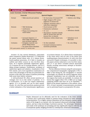
PATHOLOGY BOX 9-1
Lower Extremity Arterial Ultrasound Findings
Grayscale Spectral Doppler Color Imaging
Normal
Stenosis
Aneurysm
Occlusion
• Walls smooth and uniform
• Wall thickening
• Calcification
• Plaque encroaching into
vessel lumen
• Increased diameter 1.5 times larger than adjacent more proximal segment
• Echogenic material com- pletely filling lumen
• No focal areas of increased PSV • Slight change in PSV across
segments of arterial tree
• Triphasic waveform with re- verse flow component
• Focal increase in PSV
• Poststenotic turbulence
• Vr 2.0
• Monophasic waveform distally
with no reverse flow and con-
tinuous flow in diastole
• Turbulence within dilated area
• No flow identified
• Increased resistance noted in waveform proximally
• Uniform color filling
• Focal area of alias- ing
• Turbulence distally
• Turbulence in di- lated area
• No color filling
• No patent lumen identified PSV, peak systolic velocity; Vr, velocity ratio.
Invasive CA has several limitations, particularly when compared to duplex ultrasound: (1) it delineates the patent arterial lumen only, (2) it misses throm- bosed popliteal aneurysms, (3) it fails to visualize an outflow and inflow sources in very low-flow situa- tions, (4) it requires potentially nephrotoxic agents, (5) it requires the use of ionizing radiation, and (6) it delays prompt treatment. Furthermore, avoidance of nephrotoxic agents and radiation, visualization of low flow arteries, and a more expeditious examination are some of the advantages of DA that are particularly im- portant in this often sick subset of patients presenting with acute lower limb(s) ischemia.
A well-performed DA offers several practical ad- vantages over CA in this subset of patients: (1) it is noninvasive; (2) it does not require nephrotoxic agents; (3) it is portable and can be done expedi- tiously; (4) color flow and waveform analysis provide a better estimation of the hemodynamic significance
of occlusive disease; (5) it allows direct visualization of the entire artery and not only of the lumen thus enabling plaque characterization; (6) with color flow and power Doppler techniques, it is possible to iden- tify patent arteries subjected to very low-flow states; and (7) it can detect occluded arterial aneurysms thereby avoiding unnecessary attempts at thrombo- embolectomies.
The key difference between using DA and preopera- tive computerized tomographic angiography (CTA) or MRA is that during DA, the sonographer or vascular technologist can identify the arterial segments where adequate visualization was not achievable and alert the surgeon that results are unreliable. In contrast, the factors that suggest when CTA or MRA are no longer supplying reliable data have yet to be identified.27–30 Based on the present experience of the authors, about 90% of lower extremity revascularization procedures can be performed based on preoperative DA alone.
SUMMARY
Duplex ultrasound can be efficiently used for the evaluation of the lower extremi- ty arteries and is particularly helpful in patients being evaluated for lower extremity revascularization. It is a highly operator-dependent test that demands constant optimi- zation of the image by an operator who has mastered ultrasound technology, hemody- namics, and has a deep knowledge of pertinent anatomy. The advantages, limitations, and results of the technique are promising and may significantly improve with the use of exceptional quality imaging provided by contemporary ultrasound instruments.
