
Page 256 - Libro 2
P. 256
236
PART 4 — PERIPHERAL VENOUS
Figure 15-12 A transverse view of the axillary artery (in red) and vein (in blue) taken from the axilla. Note how there is spa- tial separation between the two vessels.
will be located on either side of the brachial artery. At this point, the brachial veins will be fairly small (Fig. 15-13).
Brachial Vein
The brachial vein is often a bifid system. The brachi- al veins will accompany the brachial artery until just below the antecubital fossa. The brachial veins are formed by the junction of two radial and two ulnar veins at the level of the antecubital fossa. The paten- cy of the brachial veins should include clear images of both brachial veins with compressed and noncom- pressed views. Some laboratories also choose to doc- ument venous spectral Doppler signals at this level.
Radial Veins
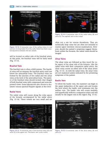
The radial veins will course along the volar aspect of the forearm accompanied by the radial artery (Fig. 15-14). These vessels are very small and are
Figure 15-13 A transverse view of the brachial artery (A) and veins (V) in the upper arm. The basilic vein is also noted.
Figure 15-14 A transverse view of the radial artery (A) and veins ( V ). The cephalic vein is also noted.
often not a site for venous thrombosis. They are technically deep veins but are often not included in routine upper extremity venous examinations. How- ever, should the patient’s symptoms suggest throm- bosis within the forearm, the radial veins should be imaged.
Ulnar Veins
The ulnar veins are followed as they travel the vo- lar aspect of the ulnar side of the forearm. Like the radial veins with their companion radial artery, the ulnar veins course on either side of the ulnar artery (Fig. 15-15). As with the radial veins, the ulnar veins are not examined unless indicated by the presenting symptoms of the patient.
Basilic Vein
To follow the basilic vein, the examiner can begin in the upper midportion of the upper arm and locates the level where the basilic vein terminates into the axillary vein. The basilic vein will course medially and superficially without a companion artery and will usually be the largest vein in the region (Fig. 15-16).
Figure 15-15 A transverse view of the ulnar artery (A) and veins ( V ).
