
Page 431 - Libro 2
P. 431
27 — Vascular Applications of Ultrasound Contrast Agents
411
AB
C
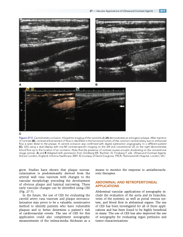
Figure 27-5 Carotid artery occlusion. A baseline imaging of the carotid bulb (A) demonstrates an echogenic plaque. After injection of contrast (B), contrast-enhancement of flow is identified in the functional lumen of the common carotid artery, but no enhanced flow is seen distal to the plaque. A carotid occlusion was confirmed with digital subtraction angiography. In a different patient (C), CES using a dual-display with low-MI contrast-specific imaging on the left and conventional US on the right demonstrates blood flow up to the location of an occlusion. Note that the presence of contrast causes acoustic shadowing on the conventional image (arrow). (A and B Adapted with permission from Goldberg BB, Raichlen JS, Forsberg F, eds. Ultrasound Contrast Agents. 2nd ed. London, England: Informa Healthcare; 2001. C Courtesy of David Cosgrove, FRCR, Hammersmith Hospital, London, UK.)
grow. Studies have shown that plaque neovas- cularization is predominantly derived from the arterial wall vasa vasorum with changes to the vascular morphology preceding the development of obvious plaque and luminal narrowing. These early vascular changes can be identified using CES (Fig. 27-7).
In the future, the use of CES for evaluating the carotid artery vasa vasorum and plaque neovascu- larization may prove to be a valuable, noninvasive method to identify patients who have vulnerable plaques and to better determine a patient’s risk of cardiovascular events. The use of CES for this application could also complement sonographic measurements of the intima-media thickness as a
means to monitor the response to antiatheroscle- rotic therapies.
ABDOMINAL AND RETROPERITONEAL APPLICATIONS
Abdominal vascular applications of sonography in- clude the evaluation of the aorta and its branches, veins of the systemic as well as portal venous sys- tem, and blood flow in abdominal organs. The use of CES has been investigated for all of these appli- cations and has been found to be highly beneficial in many. The use of CES has also improved the use of sonography for evaluating organ perfusion and tumor characterizations.
