
Page 71 - Libro 2
P. 71
4 — The Extracranial Duplex Ultrasound Examination
51
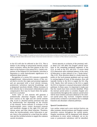
Figure 4-21 Duplex images of a patent proximal internal carotid artery and abnormal, blunted, and resistive Doppler arterial flow. Findings indicate distal extracranial versus intracranial severe stenosis or occlusion of the internal carotid artery.
in the ICA will also be reflected in the CCA. This is similar to the setting of extracranial internal carotid artery occlusion, where the flow pattern in the CCA takes on the features of the patent ECA. Careful com- parison of the bilateral ICA end-diastolic velocities is imperative to verify hemodynamic significance of a unilateral distal stenosis.
Recognition of a severe ECA stenosis is generally straightforward. Atherosclerotic lesions of this ves- sel tend to involve the origin and proximal segments and are associated with a focal velocity increase, poststenotic turbulence (spectral broadening), and a dampened waveform distally. Diffuse increases in ECA velocity due to intracranial collateralization and compensatory flow may be found when the ipsilat- eral ICA is occluded.
Aortic valve or root stenosis will generate a symmetrically abnormal Doppler arterial wave- form contour in the right and left carotid systems (Fig. 4-22A–C). Brachial systolic pressures may also be symmetrically low depending on the severity of the stenosis. Severe stenosis or occlusion of the brachiocephalic artery creates decreased pressure and waveform changes in the right CCA and sub- clavian artery and their distal branches. Therefore, when the brachiocephalic artery bifurcation is pat- ent, there is the potential for a steal from the subcla- vian and the vertebral arteries to supply the common carotid circulation.
Severe stenosis or occlusion of the proximal, mid-, or distal CCA will affect the Doppler arterial wave- form in the remaining peripheral segments of the CCA, as well as the ICA, and ECA. A severe distal CCA obstruction with continued patency of the carot- id bifurcation is often referred to as a “choke lesion” (Fig. 4-23). Flow direction distal to a choke lesion de- pends on the local pressure gradients and can result in varying degrees of steal. In this situation, flow will typically reverse in the ECA to supply the ICA (Fig. 4-24). Rarely, flow will reverse in the ICA to supply the ECA in response to unusual intracranial collateral pathways. In these cases, it is important to make sure that the vessels have been correctly identified. When the internal carotid artery is occluded, Doppler arte- rial waveforms throughout the common carotid artery will be more resistive and more identical to the exter- nal carotid artery (Fig. 4-25A,B). Complete external carotid artery occlusion is uncommon due to multiple branches and abundant collateral pathways.
Special Considerations
Low cardiac output and a poor ejection fraction can affect systemic arterial pressure and Doppler arterial waveform contour throughout the carotid system. Doppler waveforms will be dampened with delayed acceleration in every artery evaluated, yet no ste- nosis or poststenotic turbulence will be identified.
