
Page 485 - AWSAR 2.0
P. 485
cancer cells infected with S. typhimurium rp2 and find out the exact happenings and record events from the entry of S. typhimurium rp2 into cancer cells to the destruction of cancer cells. We took the help of a green fluorescent protein (GFP) gene. Once the gene was inside S. typhimurium rp2 cells, it induced the synthesis of GFP and the cells gave out green fluorescence, that is, they literally appeared green when seen under a special microscope called confocal laser scanning microscope (CLSM). CLSM facilities were provided by Dr NafisaBalasinor,NationalInstituteforResearch in Reproduction Health, Parel, Mumbai.
So for detecting and imaging the anti– cancer activity of S. typhimurium rp2, a GFP- expressing plasmid, which harbored gfp gene, was transformed into S. typhimurium rp2 by CaCl2 procedure. As GFP is synthesized by S. typhimurium rp2 cells, it is possible to visualize their growth inside cancer cells and find out the time required for killing
cancer cells, using confocal
laser scanning microscopy.
Caco-2 cells were
propagated in a six-well tissue
culture plate for detecting
the anti–cancer activity of
S. typhimurium rp2 inside
them. S. typhimurium rp2 cell
suspension was added to
Caco-2 cells and incubated
for 1, 3, 6, and 12 h. Images
were captured using the CLSM.
We expected S. typhimurium rp2 to enter into the cancer cells and kill them. As the S. typhimurium rp2 cells multiplied inside cancer cells, the amount of green fluorescence inside cancer cells also increased.
We measured fluorescence after 1, 3, 6, and 12 h using ImageJ software, which gave a plot of integrated intensity, which was quantified. After 6–12 h, it reached a plateau. There was a sharp increase in fluorescence
Ms. Rasika Pawar || 461
associated with Caco-2 cells 3 h after adding S. typhimurium rp2 cells, indicating the start of cancer cell destruction. Caco-2 cells were completely killed 12 h after S. typhimurium rp2 cells were added. When we saw this under the microscope, it was a visual treat! No words are apt and sufficient to describe that moment! What should I say Moment of joy? Moment of subjugation? Or all.
The unique aspect of this method of killing cancer cells is that it’s a way of life for Salmonella. S. typhimurium rp2 can grow under anaerobic or low-oxygen conditions. They survive and multiply ubiquitously. Cancer cells usually have a hypoxic environment around them. The major reason for this is the paucity of blood supply, which results in hypoxia. However, this, by no means, is a hindrance to the growth of Salmonella, and as a result, it colonizes both large and small tumors.
The most important problem in the treatment of cancer is reaching these imperceptible hypoxic areas with chemotherapeutic drugs as all of them require blood flow for their transport across cells and tissues. Also, radiation therapy is of little use as it also requires oxygen for its
action in cancer tissues. Colorectal cancer is the
third most common cause of cancer deaths in the world, and hence an exemplar change
in treatment is urgently needed. Research in cancer biology will always be in the pursuit of exploring panacea that would selectively identify and kill cancerous cells. Bacterial therapy of cancer is a novel paradigm changer. S. typhimurium has advantages including natural cytotoxicity, motility, and chemotaxis for killing cancer cells. Our study uses a mutagenesis strategy for the development of novel S. typhimurium rp2 as a potential
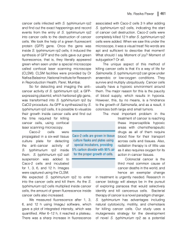
Caco-2 cells are grown in tissue culture flasks and plates using special incubators, providing 5% carbon dioxide with 95% air for the proper growth of cells.
