
Page 93 - AWSAR_1.0
P. 93
Population and Ecology Symbiosis – Wetland, Macrophyte and Fish
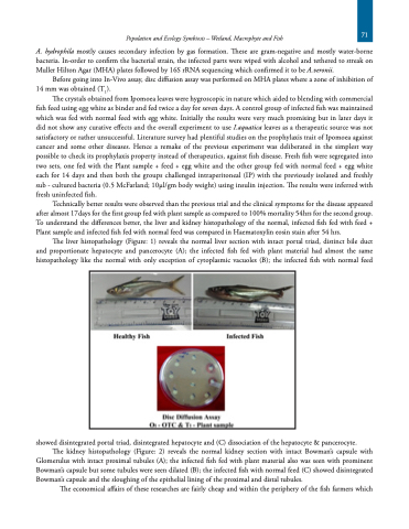
A. hydrophila mostly causes secondary infection by gas formation. These are gram-negative and mostly water-borne bacteria. In-order to confirm the bacterial strain, the infected parts were wiped with alcohol and tethered to streak on Muller Hilton Agar (MHA) plates followed by 16S rRNA sequencing which confirmed it to be A.veronii.
Before going into In-Vivo assay, disc diffusion assay was performed on MHA plates where a zone of inhibition of 14 mm was obtained (T1).
The crystals obtained from Ipomoea leaves were hygroscopic in nature which aided to blending with commercial fish feed using egg white as binder and fed twice a day for seven days. A control group of infected fish was maintained which was fed with normal feed with egg white. Initially the results were very much promising but in later days it did not show any curative effects and the overall experiment to use I.aquatica leaves as a therapeutic source was not satisfactory or rather unsuccessful. Literature survey had plentiful studies on the prophylaxis trait of Ipomoea against cancer and some other diseases. Hence a remake of the previous experiment was deliberated in the simplest way possible to check its prophylaxis property instead of therapeutics, against fish disease. Fresh fish were segregated into two sets, one fed with the Plant sample + feed + egg white and the other group fed with normal feed + egg white each for 14 days and then both the groups challenged intraperitoneal (IP) with the previously isolated and freshly sub - cultured bacteria (0.5 McFarland; 10μl/gm body weight) using insulin injection. The results were inferred with fresh uninfected fish.
Technically better results were observed than the previous trial and the clinical symptoms for the disease appeared after almost 17days for the first group fed with plant sample as compared to 100% mortality 54hrs for the second group. To understand the differences better, the liver and kidney histopathology of the normal, infected fish fed with feed + Plant sample and infected fish fed with normal feed was compared in Haematoxylin eosin stain after 54 hrs.
The liver histopathology (Figure: 1) reveals the normal liver section with intact portal triad, distinct bile duct and proportionate hepatocyte and pancerocyte (A); the infected fish fed with plant material had almost the same histopathology like the normal with only exception of cytoplasmic vacuoles (B); the infected fish with normal feed
showed disintegrated portal triad, disintegrated hepatocyte and (C) dissociation of the hepatocyte & pancerocyte.
The kidney histopathology (Figure: 2) reveals the normal kidney section with intact Bowman’s capsule with Glomerulus with intact proximal tubules (A); the infected fish fed with plant material also was seen with prominent Bowman’s capsule but some tubules were seen dilated (B); the infected fish with normal feed (C) showed disintegrated
Bowman’s capsule and the sloughing of the epithelial lining of the proximal and distal tubules.
The economical affairs of these researches are fairly cheap and within the periphery of the fish farmers which
71
