
Page 17 - CJO_F18
P. 17
REVIEW
All tissues of the ocular surface may be affected in allergic conjunctivitis. Conjunctival injection may be mild to
moderate in patients with allergic conjunctivitis, but tends to be superficial. Chemosis may seem out of propor-
tion to the degree of redness, producing a balloon effect, or may be observed only as a milky or glassy appearance
of the bulbar conjunctiva. Both may be most noticeable towards the plica semilunaris. A papillary response is
2
expected on the palpebral conjunctiva; however, it may be masked by allergy-associated chemosis. The eyelids
1
may be hyperemic or edematous, and specific observations of allergy masqueraders such as blepharitis should be
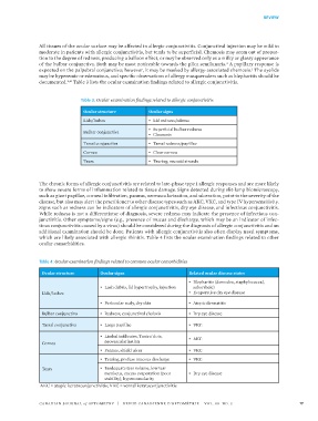
documented. Table 3 lists the ocular examination findings related to allergic conjunctivitis.
2,16
Table 3: Ocular examination findings related to allergic conjunctivitis
Ocular structure Ocular signs
Lids/lashes • Lid redness/edema
• Superficial bulbar redness
Bulbar conjunctiva
• Chemosis
Tarsal conjunctiva • Tarsal redness/papillae
Cornea • Clear cornea
Tears • Tearing, mucoid strands
The chronic forms of allergic conjunctivitis are related to late-phase type I allergic responses and are more likely
to show severe forms of inflammation related to tissue damage. Signs detected during slit-lamp biomicroscopy,
such as giant papillae, corneal infiltration, pannus, neovascularization, and ulceration, point to the severity of the
disease, but also may alert the practitioner to other disease types such as AKC, VKC, and type IV hypersensitivity.
Signs such as redness can be indicators of allergic conjunctivitis, dry eye disease, and infectious conjunctivitis.
While redness is not a differentiator of diagnosis, severe redness may indicate the presence of infectious con-
junctivitis. Other symptoms/signs (e.g., presence of mucus and discharge, which may be an indicator of infec-
tious conjunctivitis caused by a virus) should be considered during the diagnosis of allergic conjunctivitis and an
additional examination should be done. Patients with allergic conjunctivitis also often display nasal symptoms,
which are likely associated with allergic rhinitis. Table 4 lists the ocular examination findings related to other
ocular comorbidities.
Table 4: Ocular examination findings related to common ocular comorbidities
Ocular structure Ocular signs Related ocular disease states
• Blepharitis (demodex, staphylococcal,
• Lash debris, lid hypertrophy, injection seborrheic)
Lids/lashes • Evaporative dry eye disease
• Periocular scaly, dry skin • Atopic dermatitis
Bulbar conjunctiva • Redness, conjunctival chalasis • Dry eye disease
Tarsal conjunctiva • Large papillae • VKC
• Limbal infiltrates, Tantra' dots, • AKC
Cornea neovascularisation
• Pannus, shield ulcer • VKC
• Tearing, profuse mucous discharge • VKC
Tears • Inadequate tear volume, low tear
meniscus, excess evaporation (poor • Dry eye disease
stability), hyperosmolarity
AKC = atopic keratoconjunctivitis; VKC = vernal keratoconjunctivitis
CANADIAN JOURNAL of OPTOMETRY | REVUE CANADIENNE D’OPTOMÉTRIE VOL. 80 NO. 3 17
38668_CJO_F18 August 10, 2018 8:58 AM APPROVAL: ___________________ DATE: ___________________