
Page 30 - DMX HANDBOOK 4TH EDITION
P. 30
24. Repeat MRIs can objectively prove accelerated degeneration.
We have known for many years that CAD injury predisposes the victims to accelerated degeneration. Previously, that concept has
only been a hypothetical one as it relates to any given patient/client. Recently, however, the lawyers at the Schmidt Salita Law Firm
have discovered that, in some cases, there is objective, irrefutable proof of highly accelerated degeneration in some of the clients
who have been found on DMX to have significant ligamentous laxity. One of those cases is presented below. In “Gina’s Case,” a 14-
year-old, previously healthy girl was found to have the equivalent of 30 years of accelerated degeneration that had occurred in only 2
years!!!
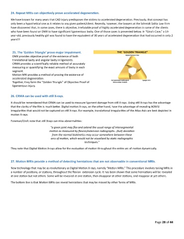
25. The 'Golden Triangle' prove major impairment.
DMX provides objective proof of the existence of both
translational laxity and angular laxity in ligaments.
CRMA provides a scientifically reliable method of accurately
measuring or quantifying the exact amount of laxity in each
segment.
Motion MRI provides a method of proving the existence of
accelerated degeneration.
Together, they form the “Golden Triangle” of Objective Proof of
ligamentous injury.
26. CRMA can be used with still X-rays.
It should be remembered that CRMA can be used to measure ligament damage from still X-rays. Using still X-rays has the advantage
that the clarity of the film is much better. Digital motion X-rays, on the other hand, have the advantage of revealing AOMSI
irregularities that would not be captured on still X-rays. For example, translational irregularities of the Atlas-Axis are best depicted in
motion X-rays.
Foreman/Croft note that still Xrays can miss abnormalities:
“a given joint may flex and extend the usual range of intersegmental
motion as measured by flexion/extension radiographs…(but) deviation
from the normal biokinetics may occur somewhere between these
arcs of motion, which would not be visualized by static radiographic
techniques.”
They note that Digital Motion X-rays allow for the evaluation of motion throughout the entire arc of motion dynamically.
27. Motion MRIs provide a method of detecting herniations that are not observable in conventional MRIs.
New technology that may be as revolutionary as Digital Motion X-rays, namely “Motion MRIs.” This procedure involves taking MRIs in
a number of positions, or stations, throughout the flexion- extension cycle. It has been shown that some herniations will be revealed
at one station but not others. Some will be revealed at one station, then disappear at other stations, and reappear at yet others.
The bottom line is that Motion MRIs can reveal herniations that may be missed by other forms of MRIs.
Page 28 of 44