
Page 58 - Chiron Calling Autum 2021/Spring 2022
P. 58
important, as well as always trying to find and provide shade for the dogs as often as we could. It was the first time I had seen dogs actively searching for shade in a person’s shadow, MWD Twitter did this a lot. After spending an hour on an open TCV, MWD Etna had become very lethargic and her handler thought that she did not seem her normal self. I quickly did a TPR which showed a high heart rate and a temperature of 39.9 in addition to her panting. This showed us she was suffering from the heat, so we had
to act quickly to prevent her from becoming a casualty. Thankfully there was a MAN truck with air con running, so we placed her in the cab and used surgical spirit on her foot pads to encourage cooling. I had to make the decision to pull her off
the rest of the patrol as she was not physically able to carry on at this point without causing further harm.
After the exercise had finished, we had the opportunity to go back out for 6 days where we were able to conduct dog training, as well
as go on a pistol range. I took this opportunity to Army Medics how to perform veterinary skills on a dog that may go down on patrol. It was interesting to see how similar their human drills are to our veterinary ones.
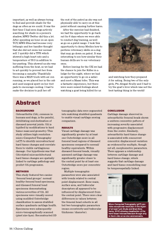
Before leaving for the UK we had the chance to join the Rifles in a lodge for the night, where we had an opportunity to go on a safari and meet a Masai tribe. This was
a fantastic experience, but there were more mixed feelings about watching a goat being killed for us
and watching how they prepared
it for eating. Being two of the only girls, Pte Abigail Scully and I had to try the goat’s liver which was not the best tasting thing in the world!
Abstract
BACKGROUND
Osteoarthritis (OA), common in humans and dogs, is the painful, debilitating end-destination of diseased synovial joints. OA is typified by modifications to hard tissue mass and geometry. This study utilises high resolution micro-Computed Tomography (uCT) to identify microstructural hard tissue changes and correlate them to visible cartilaginous damage. Our hypothesis was that OA-related microarchitectural hard tissue changes are spatially linked to cartilage pathology and predict OA progression.
METHODS
This study featured two canine ‘femoral head groups’: normal/ healthy femoral head specimens; and diseased femoral head specimens demonstrating
various severities of OA. All specimens were visually scored using modified Outerbridge classification to assess stratified surface quadrants cartilage health. Specimens were subsequently micro-tomographically scanned (pixel size 9μm). Reconstructed 3D
tomographic data were segmented into matching stratified quadrants to enable visual cartilage scoring comparison.
RESULTS
Visual cartilage damage was significantly greater by at least one Outerbridge score in all femoral head regions of diseased specimens compared to normal/ healthy equivalents. Within diseased femoral heads, visually assessed cartilage damage was significantly greater closer to
the central point by at least one Outerbridge score per concentric region.
Multiple tomographic parameters were also associated with trends related to central point displacement. Bone mass, surface area, and trabecular descriptors all appeared to be influenced by displacement from the central point. There were clear differences in values between
the femoral head cohorts in all but two tomographic parameters (mean eccentricity and trabecular thickness / diameter).
CONCLUSION
Cartilage damage observed in osteoarthritic femoral heads shows a uniform concentric pattern of increasing severity associated
with progressive displacement from the centre. Similarly, osteoarthritic hard tissue change is associated with concurrent concentric displacement variation as evidenced by multiple, though not all, morphometric parameters. There appears a relationship between cartilage damage and hard tissue change, which suggests that cartilage damage and hard tissue remodelling may be temporospatially linked.
Micro-Computed Tomography (μCT) pro- vides exceptional resolution in the region of at least 1μm (Huda and Brad Abra- hams, 2015) allowing visualisation of the hard tissue microarchitecture though only in the in vitro or ex vivo environments.
56 / Chiron Calling
