
Page 18 - November2020
P. 18
Important Moments in the History of CT
1900 – Italian radiologist Alessandro Vallebona invented tomography, using radiographic film to see
a single slice of the body.
1960 – Increased power and availability of computers sparked new research to create practical
computational tomographic images.
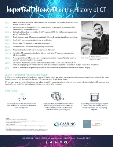
1967 – Sir Godfrey Hounsfield invented the first CT scanner at EMI Central Research Laboratories
using X-ray technology.
1971 – The first patient brain CT was performed in Wimbledon, England and publicized a year later.
1973 – The first CT scanners are installed in the United States.
1980 – Three million CT examinations are being performed.
1990s – Portable/mobile CT scanners begin growing in popularity.
2005 – The annual number of CT examinations grows to 68 million.
2007 – Half of the CT scanners installed in the U.S. are multi-slice CT scanners with more than
64-slice capability. Sir Godfrey Hounsfield.
2008 – A new generation of CT scanners are developed and can take images of beating hearts or (Credit: http://www.britannica.com/biography/Godfrey-
coronary arteries in less than one second. Newbold-Hounsfield)
2009 – Dr. Mathias Prokop discusses the clinical implications of the 16-cm wide detector CT. The
wider coverage per gantry rotation enables more dynamic scanning and the ability to do multiple acquisitions in less time.
2010 – The FDA launches its Image Wisely initiative to reduce unnecessary radiation exposure from medical imaging.
About Computed Tomography Exams
A CT scan combines a series of X-ray images taken at different angles and uses a computer to create cross-sectional images (slices) of the bones,
blood vessels and soft tissues inside the body. CT scans are more detailed than X-rays.
CT scans are used for many different uses but is best for quickly examining patients who may have sustained internal injuries. CT scans are used
to visualize any part of the body, diagnose diseases and planning for treatment.
Fast Facts:
It is widely rumored that the Beatles’ record Adaptive Statistical, model-based and hybrid- 3D printing allows radiologists to print
sales in the 1960’s helped fund the first CT iterative image reconstruction techniques CT data in three dimensions, assisting
scan’s development. contributed to a 70-80% radiation dose in surgical planning and prototyping
reduction over the past four years. implants.
Sources:
International Society for Computed Tomography
https://www.isct.org/computed-tomography-blog/2017/2/10/half-a-century-in-ct-how-
computed-tomography-has-evolved
Science Learning Hub
https://www.sciencelearn.org.nz/resources/1906-developments-in-medical-imaging-timeline
Mayo Clinic
https://www.mayoclinic.org/tests-procedures/ct-scan/about/pac-20393675#targetText=A%20
computerized%20tomography%20(CT)%20scan,soft%20tissues%20inside%20your%20body.
800-228-5462 | 402-334-5000 | www.cassling.com | 13808 F Street | Omaha, NE 68137