
Page 204 - C:\Users\uromn\Videos\seyyedi pdf\
P. 204
Abdollahi, et al.: Antibacterial effects of 940 nm diode laser
under 5% CO between each inoculation at 37°C, and
2
the entire inoculation and incubation period lasted
1 week. After the last inoculation and 48 h, sampling
was performed from all the root canals to determine
colony forming units counts. The inoculation process
was carried out before each intervention and after
the procedure. A #60 paper point was inserted into
each root canal for sampling. Immediately, on a plate
containing blood agar, four concentric circles were
drawn that decreased in diameter toward the center
with a paper cutter. The plates were incubated at
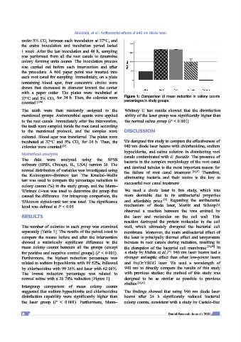
37°C and 5% CO for 24 h. Then, the colonies were Figure 1: Comparison of mean reduction in colony counts
2
counted. [8,26] percentages in study groups.
The teeth were then randomly assigned to the Whitney U test results showed that the disinfection
mentioned groups. Antimicrobial agents were applied ability of the laser group was significantly higher than
to the root canals. Immediately after the intervention, the normal saline group (P < 0.001).
the teeth were sampled inside the root canal according
to the mentioned protocol, and the samples were DISCUSSION
cultured. Blood agar was transferred. The plates were
incubated at 37°C and 5% CO for 24 h. Then, the We designed this study to compare the effectiveness of
2
colonies were counted. [26] 940 nm diode laser beams with chlorhexidine, sodium
Statistical analysis hypochlorite, and saline solution in disinfecting root
canals contaminated with E. faecalis. The presence of
The data were analyzed using the SPSS bacteria in the complex morphology of the root canal
software (SPSS, Chicago, IL, USA) version 26. The and dentinal tubules is the most important reason for
normal distribution of variables was investigated using the failure of root canal treatment. [12,27] Therefore,
the Kolmogorov–Smirnov test. The Kruskal–Wallis eliminating bacteria and their toxins is the key to
test was used to compare the percentage reduction in successful root canal treatment.
colony counts (%) in the study group, and the Mann–
Whitney U‑test was used to determine the group that We used a diode laser in this study, which was
caused the difference. For intragroup comparison, the more desirable due to its antibacterial properties
[21]
Wilcoxon signed‑rank test was used. The significance and affordable price. Regarding the antibacterial
[28]
level was defined at P < 0.05. mechanism of diode laser, Moritz and Schoop
observed a reaction between the ions emitted by
RESULTS the laser and molecules on the cell wall. This
reaction destroyed the protein molecules in the cell
The number of colonies in each group was examined wall, which ultimately disrupted the bacterial cell
separately [Table 1]. The results of the paired t‑test to membrane. Moreover, the main antibacterial effect of
compare the means before and after the intervention the laser is principally thermal effect and temperature
showed a statistically significant difference in the increase in root canals during radiation, resulting in
mean colony counts between all the groups (except the disruption of the bacterial cell membrane. [22,29] In
for positive and negative control groups) (P < 0.001). a study by Mehta et al., 940 nm laser beams had a
[30]
Furthermore, the highest reduction percentage was stronger antiseptic effect than other low‑power lasers
related to sodium hypochlorite with 99.52%, followed and Er,Cr:YSGG laser. We used a wavelength of
by chlorhexidine with 99.36% and laser with 62.06%. 940 nm to directly compare the results of this study
The lowest reduction percentage was related to with previous studies; the method of this study was
normal saline with a 26.78% reduction [Figure 1]. designed to be as similar as possible to previous
Intergroup comparison of mean colony counts studies. [26,31]
suggested that sodium hypochlorite and chlorhexidine The findings showed that using 940 nm diode laser
disinfection capability were significantly higher than beams after 24 h significantly reduced bacterial
the laser group (P < 0.001). Furthermore, Mann– colony counts, consistent with a study by Castelo‑Baz
4 Dental Research Journal / 2024