
Page 14 - DP Vol 21 No1_Neat
P. 14
AESTHETIC DENTISTRY SECTION
HUMAN TAKE ON THE DIGITAL
CONUNDRUM
Arshya Gandhi
INTRODUCTION
The arrival of digitally-driven technology has, indeed,
revolutionized aesthetic dentistry and opened up a world
where precision can be heightened while meeting individual
patient needs. Digital Smile Design (DSD) is the most recent
tool for visualizing and robotically planning dental aesthetics.
The following case report describes a 25-year-old male patient
affected by hypoplastic discoloration and generalized maxillary
anterior spacing, (Figure 1a) seeking an aesthetic solution.
The treatment of choice for this particular clinical situation
was Emax veneers, ensuring a quick and successful resolution,
supported by the DSD protocol.
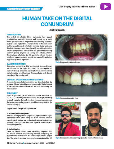
Fig 1a: Pre-operative intraoral image
CASE PRESENTATION
The patient came with a chief complaint of white and brown
discoloration on the upper front teeth (11, 21) (Figure 1b)
since childhood, along with spacing between all the anterior
teeth, including a midline space. The mandibular arch showed
crowding of the anterior teeth.
CLINICAL EXAMINATION AND DIAGNOSIS
A comprehensive clinical evaluation was done including the
extraoral (Figure 1c) and intraoral photographs. Digital models
of the dentition were fabricated via intraoral scans using the
iTero scanner.
TREATMENT
Tooth Preparation: The six maxillary anterior teeth (13, 12,
11, 21, 22, 23) were prepared for Emax veneer placement by Fig 1b: Pre-operative incisal view
gradually reducing the tooth structure to ensure adequate space
for each corresponding veneer type, without compromising the
structural integrity.
Digital Smile Design (DSD) Protocol
1. Scanning and Data Upload
After the tooth preparation (Figure 2a), high-resolution digital
impressions were taken using the iTero intraoral scanner,
recording each contour and surface detail required for treatment
planning. The digital files were then imported into the exocad
software (Figure 2b).
2. Smile Creation
Once the digital models were successfully imported into
the exocad software, the next step involved integrating the
patient’s facial features into the smile design process (Figure
3b). The patient’s extraoral photographs were uploaded for 3D Fig 1c: Pre-operative extraoral image (note the underconfident smile)
14 Dental Practice I January-February 2025 I Vol 21 No 1