
Page 26 - Dental Practice Vol 17 No.5_
P. 26
periodontic section
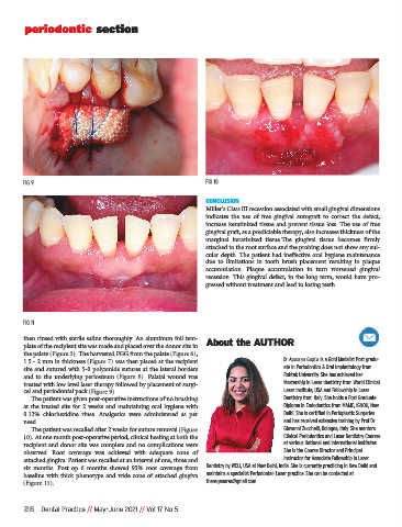
FIG 9 FIG 10
CONCLUSION
Miller’s Class III recession associated with small gingival dimensions
indicates the use of free gingival autograft to correct the defect,
increase keratinized tissue and prevent tissue loss. The use of free
gingival graft, as a predictable therapy, also increases thickness of the
marginal keratinized tissue.The gingival tissue becomes firmly
attached to the root surface and the probing does not show any sul-
cular depth. The patient had ineffective oral hygiene maintenance
due to limitations in tooth brush placement resulting in plaque
accumulation. Plaque accumulation in turn worsened gingival
recession. This gingival defect, in the long term, would have pro-
gressed without treatment and lead to losing teeth.
FIG 11
then rinsed with sterile saline thoroughly. An aluminum foil tem-
plate of the recipient site was made and placed over the donor site in About the AUTHOR
the palate (Figure 5). The harvested FGG from the palate (Figure 6),
1.5 - 2 mm in thickness (Figure 7) was then placed at the recipient Dr Apoorva Gupta is a Gold Medalist Post gradu-
site and sutured with 5-0 polyamide sutures at the lateral borders ate in Periodontics & Oral Implantology from
and to the underlying periosteum (Figure 8). Palatal wound was Rohtak University. She has achieved her
treated with low level laser therapy followed by placement of surgi- Mastership in Laser dentistry from World Clinical
cel and periodontal pack (Figure 9). Laser Institute, USA and Fellowship in Laser
The patient was given post-operative instructions of no brushing Dentistry from Italy. She holds a Post Graduate
at the treated site for 2 weeks and maintaining oral hygiene with Diploma in Endodontics from MAMC, IGNOU, New
0.12% chlorhexidine rinse. Analgesics were administered as per Delhi. She is certified in Perioplastic Surgeries
need. and has received extensive training by Prof Dr
The patient was recalled after 2 weeks for suture removal (Figure Giovanni Zucchelli, Bologna, Italy. She mentors
10). At one month post-operative period, clinical healing at both the Clinical Periodontics and Laser Dentistry Courses
recipient and donor site was complete and no complications were at various National and International Institutes.
observed. Root coverage was achieved with adequate zone of She is the Course Director and Principal
attached gingiva. Patient was recalled at an interval of one, three and Instructor for Associate Fellowship in Laser
six months. Post op 6 months showed 95% root coverage from Dentistry by WCLI, USA at New Delhi, India. She is currently practicing in New Delhi and
baseline with thick phenotype and wide zone of attached gingiva maintains a specialist Periodontal- Laser practice. She can be contacted at
(Figure 11). theregeneras@gmail.com
26 Dental Practice // May-June 2021 // Vol 17 No 5