
Page 55 - Dental Practice Vol 17 No.5_
P. 55
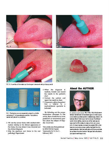
FIG 3-6: A portion of the lesion on the tongue is removed using a biopsy punch.

                                                                                   About the AUTHOR
                                                     5. When  the  diagnosis  is
                                                       received,  transfer  and  record
                                                       the  results  in  the  patient’s
                                                       chart.
                                                     6. Contact  the  patient  and
                                                       report the results to them.
                                                     7. If necessary, advise the patient
                                                       that   a   referral   to   a
                                                       medical/dental  specialist  is
                                                       indicated.

                                                       By  following  out  the  proper  Dr. Lawrence (Larry) Gaum, DDS, FADSA, FICD, FADI, Diplomat
              FIG 7: The lesions are now separately placed in a bottle
             containing 1.7 % formaldehyde solution. The bottle is  instructions  discussed  in  this  National Dental Board of Anesthesiology is an Oral Surgeon
             sealed and placed in a container.       article, there should be no more  who maintains a private practice in Mississauga, Ontario. His
                                                     guesswork or unanswered ques-  textbook titled “Oral Surgery for the General Practitioner”,
                                                     tion  about  the  subject  of  biop-  now in its 2nd edition, remains one of the most popular
             3. Fill out the correct forms with detailed infor-  sies. Good luck.  resources for GP's looking to enhance their oral surgery
               mation  relating  to  the  clinical  appearance  of                 skills. Dr. Gaum has also produced two surgical DVDs which
               the tissue, the area where it was removed and  This article was first published  have also received enormous positive feedback from the
               the clinical diagnosis.               in SPECTRUM Dental            dental profession. Both the textbook and DVDs are available
             4. Ship  the  specimen  and  forms  to  the  oral  Teamwork,Vol.14 No.1 -  for purchase from the Spectrum Dialogue Online Bookstore
               pathology Department.                 February 2021                 (www.spectrumdialogue.com)

                                                                         Dental Practice // May-June  2021 // Vol 17 No 5 55