
Page 21 - DP Vol 17 No 4 ok
P. 21
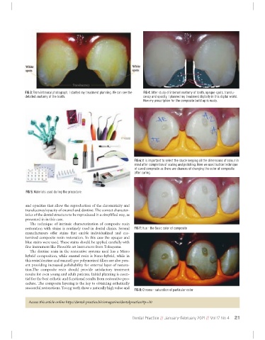
FIG 3: From intraoral photograph, I started my treatment planning. We can see the  FIG 4: After study of internal anatomy of tooth, opaque spots, translu-
             detailed anatomy of the tooth.                             cency and opacity, I planned my treatment digitally in this digital world.
                                                                        Now my prescription for the composite build up is ready.


                                                                   FIG 6: It is important to select the shade keeping all the dimensions of colour in
                                                                   mind after completion of scaling and polishing. Here we used button technique
                                                                   of cured composite as there are chances of changing the color of composite
                                                                   after curing.


              FIG 5: Materials used during the procedure.


              and opacities that allow the reproduction of the chromaticity and
              translucency/opacity of enamel and dentine. The correct character-
              istics of the dental structure to be reproduced in a simplified way, as
              presented in in this case.
                The  technique  of  intrinsic  characterization  of  composite  resin
              restoration  with  stains  is  routinely  used  in  dental  clinics.  Several  FIG 7: Hue - the Basic color of composite
              manufacturers  offer  stains  that  enable  individualized  and  cus-
              tomized composite resin restoration. In this case the opaque and
              blue stains were used. These stains should be applied carefully with
              fine instrument like Flowable art instrument from Tokuyama.
                The  dentine resin  in the  restorative  systems used  has  a  Micro
              hybrid composition, while enamel resin is Nano-hybrid, while in
              this resin(dentine and enamel) pre polymerized fillers are also pres-
              ent providing increased polishability for external layer of restora-
              tion.The  composite  resin  should  provide  satisfactory  treatment
              results for even young and adult patients. Initial planning is essen-
              tial for the best esthetic and functional results from restorative pro-
              cedure. The composite layering is the key to obtaining esthetically
              successful restorations. Young teeth show a naturally high value and
                                                                   FIG 8: Chroma - saturation of particular color

               Access this article online http://dental-practice.biz/emagazine/dentalpractice/#p=20


                                                                  Dental Practice // January-February 2021 // Vol 17 No 4  21