
Page 39 - DP Vol 17 No 4 ok
P. 39
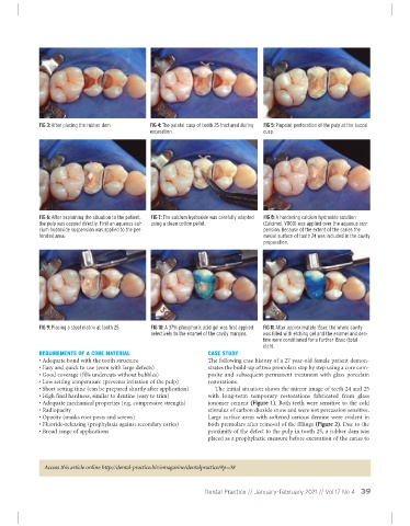
FIG 3: After placing the rubber dam.  FIG 4: The palatal cusp of tooth 25 fractured during  FIG 5: Pinpoint perforation of the pulp at the buccal
                                                 excavation.                         cusp.


              FIG 6: After explaining the situation to the patient,  FIG 7: The calcium hydroxide was carefully adapted  FIG 8: A hardening calcium hydroxide solution
             the pulp was capped directly. First an aqueous cal-  using a clean cotton pellet.    (Calcimol, VOCO) was applied over the aqueous sus-
             cium hydroxide suspension was applied to the per-                       pension. Because of the extent of the caries the
             forated area.                                                           mesial surface of tooth 24 was included in the cavity
                                                                                     preparation.


              FIG 9: Placing a steel matrix at tooth 25.  FIG 10: A 37% phosphoric acid gel was first applied  FIG 11: After approximately 15sec the whole cavity
                                                 selectively to the enamel of the cavity margins.  was filled with etching gel and the enamel and den-
                                                                                     tine were conditioned for a further 15sec (total
                                                                                     etch).
              REQUIREMENTS OF A CORE MATERIAL                      CASE STUDY
              • Adequate bond with the tooth structure             The following case history of a 27 year-old female patient demon-
              • Easy and quick to use (even with large defects)    strates the build-up of two premolars step by step using a core com-
              • Good coverage (fills undercuts without bubbles)    posite  and  subsequent  permanent  treatment  with  glass  porcelain
              • Low setting temperature (prevents irritation of the pulp)  restorations.
             • Short setting time (can be prepared shortly after application)  The initial situation shows the mirror image of teeth 24 and 25
             • High final hardness, similar to dentine (easy to trim)  with  long-term  temporary  restorations  fabricated  from  glass
             • Adequate mechanical properties (e.g. compressive strength)  ionomer cement (Figure 1). Both teeth were sensitive to the cold
             • Radiopacity                                         stimulus of carbon dioxide snow and were not percussion sensitive.
             • Opacity (masks root posts and screws)               Large surface areas with softened carious dentine were evident in
             • Fluoride-releasing (prophylaxis against secondary caries)  both premolars after removal of the fillings (Figure 2). Due to the
             • Broad range of applications                         proximity of the defect to the pulp in tooth 25, a rubber dam was
                                                                   placed as a prophylactic measure before excavation of the caries to


               Access this article online http://dental-practice.biz/emagazine/dentalpractice/#p=38


                                                                  Dental Practice // January-February 2021 // Vol 17 No 4 39