
Page 24 - Dental Practice Vol 17 No.5_Neat
P. 24
periodontic section
MANAGEMENT OF GINGIVAL RECESSION
AND LACK OF KERATINISED TISSUE
AROUND MANDIBULAR INCISORS WITH
FREE GINGIVAL GRAFT
APOORVA GUPTA
Gingival recession is a mucogingival defect that creates an esthet-
ic problem, fear of tooth loss, dentin hypersensitivity, root caries
or cervical wear. Frequently associated with questionable prog-
nosis, it is most commonly seen in the lower anterior area.Free
soft tissue autogenous graft (FGG) is a long-standing procedure
in such situation used to achieve clinical endpoints including
increasing the width of keratinized and attached gingiva, deep-
ening the vestibular depth, dissipating muscle and frenulum
pull, covering exposed root surfaces, and converting a thin peri-
odontal phenotype to a thick phenotype. FGG procedure is not
the gold standard for root coverage however the occurrence of
creeping attachment describes coronal movement of gingival
margin.
CASE PRESENTATION
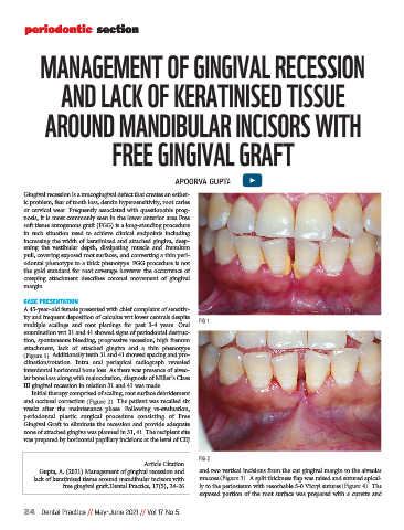
A 45-year-old female presented with chief complaint of sensitiv-
ity and frequent deposition of calculus wrt lower centrals despite FIG 1
multiple scalings and root planings for past 3-4 years. Oral
examination wrt 31 and 41 showed signs of periodontal destruc-
tion, spontaneous bleeding, progressive recession, high frenum
attachment, lack of attached gingiva and a thin phenotype
(Figure 1). Additionally teeth 31 and 41 showed spacing and pro-
clination/rotation. Intra oral periapical radiograph revealed
interdental horizontal bone loss. As there was presence of alveo-
lar bone loss along with malocclusion, diagnosis of Miller’s Class
III gingival recession in relation 31 and 41 was made.
Initial therapy comprised of scaling, root surface debridement
and occlusal correction (Figure 2). The patient was recalled six
weeks after the maintenance phase. Following re-evaluation,
periodontal plastic surgical procedure consisting of Free
Gingival Graft to eliminate the recession and provide adequate
zone of attached gingiva was planned in 31, 41. The recipient site
was prepared by horizontal papillary incisions at the level of CEJ
FIG 2
Article Citation
Gupta, A. (2021) Management of gingival recession and and two vertical incisions from the cut gingival margin to the alveolar
lack of keratinised tissue around mandibular incisors with mucosa (Figure 3). A split thickness flap was raised and sutured apical-
free gingival graft.Dental Practice, 17(5), 24-26 ly to the periosteum with resorbable 5-0 Vicryl sutures (Figure 4). The
exposed portion of the root surface was prepared with a curette and
24 Dental Practice // May-June 2021 // Vol 17 No 5