
Page 21 - Dental Practice Vol 17 No.5_Neat
P. 21
on a maxillary anterior tooth where the proximal contact and
incised edge position were developed using the Fusion Anterior
Matrix System.
CASE REPORT
A 74 year old male presented with an old class III composite on
the distal portion of his left lateral incisor (Figure 1). Recurrent
decay was noted both visually as well as radiographically.
Treatment options were discussed with patient and it was decid-
ed we would replace the restoration with a new direct compos-
ite restoration. Small amounts of composite were placed on to
the tooth and light cured to get an idea of what shade or shades
would be utilized.
The patient was anesthetized with 1/2 carpule of 4%
Articaine (Septodont) with 1:100,000 epinephrine. Isolation
was obtained with a Comfort View® Lip & Cheek Retractor
(Premier Dental). A pear shaped diamond bur (Meisinger) was
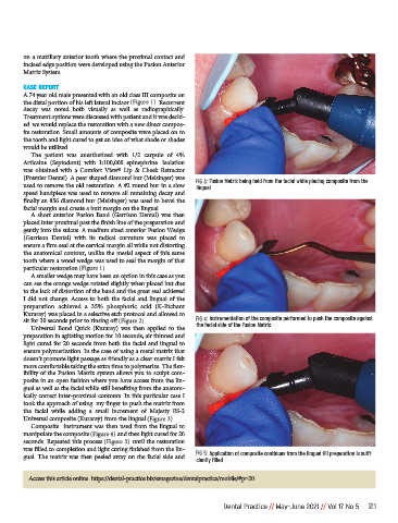
FIG 3: Fusion Matrix being held from the facial while placing composite from the
used to remove the old restoration. A #2 round bur in a slow lingual.
speed handpiece was used to remove all remaining decay and
finally an 856 diamond bur (Meisinger) was used to bevel the
facial margin and create a butt margin on the lingual.
A short anterior Fusion Band (Garrison Dental) was then
placed inter proximal past the finish line of the preparation and
gently into the sulcus. A medium sized anterior Fusion Wedge
(Garrison Dental) with its radical curvature was placed to
ensure a firm seal at the cervical margin all while not distorting
the anatomical contour, unlike the mesial aspect of this same
tooth where a wood wedge was used to seal the margin of that
particular restoration (Figure 1).
A smaller wedge may have been an option in this case as you
can see the orange wedge rotated slightly when placed but due
to the lack of distortion of the band and the great seal achieved
I did not change. Access to both the facial and lingual of the
preparation achieved a 35% phosphoric acid (K-Etchant
Kuraray) was placed in a selective etch protocol and allowed to
sit for 30 seconds prior to rinsing off (Figure 2). FIG 4: Instrumentation of the composite performed to push the composite against
Universal Bond Quick (Kuraray) was then applied to the the facial side of the Fusion Matrix
preparation in agitating motion for 10 seconds, air thinned and
light cured for 20 seconds from both the facial and lingual to
ensure polymerization. In the case of using a metal matrix that
doesn’t promote light passage as friendly as a clear matrix I felt
more comfortable taking the extra time to polymerize. The flex-
ibility of the Fusion Matrix system allows you to sculpt com-
posite in an open fashion where you have access from the lin-
gual as well as the facial while still benefiting from the anatom-
ically correct inter-proximal contours. In this particular case I
took the approach of using my finger to push the matrix from
the facial while adding a small increment of Majesty ES-2
Universal composite (Kuraray) from the lingual (Figure 3).
Composite instrument was then used from the lingual to
manipulate the composite (Figure 4) and then light cured for 20
seconds. Repeated this process (Figure 5) until the restoration
was filled to completion and light curing finished from the lin-
gual. The matrix was then peeled away on the facial side and FIG 5: Application of composite continues from the lingual till preparation is suffi-
ciently filled.
Access this article online https://dental-practice.biz/emagazine/dentalpractice/mobile/#p=20
Dental Practice // May-June 2021 // Vol 17 No 5 21